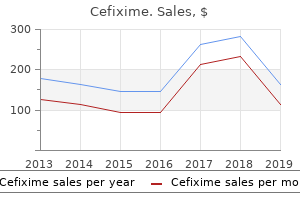
"Buy line cefixime, antibiotics for acne during pregnancy."By: Dawn Sowards Brezina, MD - Assistant Professor of Medicine
 https://medicine.duke.edu/faculty/dawn-sowards-brezina-md
Cheap cefixime online visaAs described above, acute lesions may cause focal expan sion of the wire and enhance with contrast, while persistent lesions are likely to produce atrophy. It ought to be confused that foci of periventricular T2 hyperintensity are observed with a wide range of pathologic processes and even in normal persons, particularly older ones. The identical lack of specificity of cerebral lesions pertains to those in the spinal cord. Certainly, the illness is in all probability going when one of the usual syndromes, such as optic neuritis, bilateral brainstem symptoms, or transverse myelitis, occurs in a youthful individual. However, the time-honored-and still valid-criteria for diagnosis proposed by McAlpine and colleagues (1972), requiring several lesions that were "separated in time and house," have been broadened significantly by the flexibility to detect demyelinating lesions by nonclinical means. These include visible, auditory, and somatosensory-evoked responses and the much less standardized and sometimes tested perceptual delay on visual stimulation; electro oculography; altered blink reflexes; and a change in flicker fusion of visual images. Some patients will have an entire medical remission after the initial assault, or, there could also be a collection of exacerbations, every with com plete remission; hardly ever, such exacerbations could also be severe enough to have caused quadriplegia and pseudobulbar palsy. A further 20 % relapsed in 5 to 9 years, and one other 10 p.c in 10 to 30 years. Not solely the length of this interval is remarkable, but in addition the truth that the basic pathologic course of can stay doubtlessly active for such a very long time. Perhaps not surprisingly, they found that a excessive degree of disability, as measured by the Kurtzke Disability Status Scale, was reached earlier in sufferers with the next variety of attacks, a shorter first interattack interval, and a shorter time to reach a state of reasonable incapacity. Kurtzke had earlier reported that the characteristic most predictive of long-term incapacity was the degree of disability at 5 years from the first symptom. The common relapse price in estab lished cases declines in each trimester, reaching a stage lower than one-third of the anticipated rate by the third trimester. However, there seems to be an elevated danger of exacer bations, as a lot as twofold, within the first few months postpartum (Birk and Rudick). An intensive examine of 269 pregnancies by Confavreux and colleagues (1998) established a rate of relapse of zero. A small number of sufferers die inside several months or years of the onset, but the average duration of the unwell ness is in excess of 30 years. At the top of 25 years, one-third of the surviving sufferers were nonetheless working and two-thirds had been nonetheless ambulatory (Percy et al). Other statistical analyses have given a less optimistic prognosis; these were reviewed by Matthews. Patients with delicate and quiescent forms of the disease are, in fact, much less more doubtless to be included in such surveys. Although exceptional, certainly one of our sufferers relapsed and developed massive brainstem demyelination and coma after 30 years (confirmed by postmortem examination) and instances of an aggressive myelopathy that appears after years are well-known. No environmental, dietary, or activity-related modifications are identified to alter the course of the sickness. Extensive brainstem demyelin ation of subacute evolution, involving tracts and cranial nerves sequentially, may be mistaken for a pontine glioma. The lesions may be small and single, a number of, or confluent in large regions (Akasbi). Nevertheless a number of the lesions characterize small zones of infarct necrosis quite than demyelination and are traceable to small-vessel occlusion. Others may be auto immune and demyelinating and this group of processes that have an effect on the cerebral white matter stays tough to understand. In a couple of instances, inflammatory demyelin ation with out vascular adjustments could also be seen. The distinction could also be significantly troublesome in rare instances of the vasculitic process by which the neurologic manifestations take the form of a relapsing or steroid-responsive myelitis. The distinguishing options of Beh<;et disease are recurrent iridocyclitis and meningitis, mucous mem brane ulcers of mouth and genitalia, and signs of articular, renal, lung, and multifocal cerebral disease. The persistent types of brucellosis in the Mediterranean regions and Lyme borreliosis throughout North America and Europe may cause myelopathy or encephalopathy with a number of white matter lesions on imaging studies, but in every case the historical past and other options of the illness help to identify the infectious sickness (see Chap. Such sufferers require cautious analysis for the presence of spinal cord compression from neoplasm or cervical spondylosis. As a general rule, loss of belly reflexes, erectile dysfunction, and disturbances of blad der function happen early in the midst of demyelinating myelopathy but late or under no circumstances in cervical spondylosis. A particular drawback arises when imaging procedures reveal a regional swelling of the spinal twine suggestive of a tumor. In a patient with this discovering and a subacute, saltatory myelopathy restricted to several adjacent levels (usually thoracic), a search for an arteriovenous mal formation or fistula may be required. In a number of of our patients, this discovering has led to an ill-advised try at spinal twine biopsy. A subpial sample of enhancement with gadolinium is useful in identifying sarcoid. Platybasia and basilar impression of the skull must also be considered within the differential diagnosis, however patients with these conditions normally have a characteris tic shortening of the neck; images of the bottom of the cranium are diagnostic. In every of those situations, a solitary, stra tegically positioned lesion might give rise to quite lots of neuro logic symptoms and indicators referable to the lower brainstem and cranial nerves, cerebellum, and higher cervical twine, giving the impression of dissemination of lesions. The latter are usually distin guished by their familial incidence and other associated genetic traits; by their insidious onset and gradual, regular development; and by their relative syrrunetry and stereo typed clinical sample. Intactness of stomach reflexes and sphincter function and the presence of pes cavus, kyphoscoliosis, and cardiac illness are different features that favor the diagnosis of a heredodegenerative disorder (see Chap. The many therapeutic trials of latest years, utilizing primarily anti-inflammatory and irrununosuppres sive are summarized beneath. Therefore, as mentioned earlier, therapy should be guided by the nature of the illness in each particular person and with consideration of the unwanted effects and dangers of each of the increasing group of available therapies. Corticosteroids Under the affect of corticoste roids, restoration from an acute assault, together with an attack of optic neuritis, seems to be hastened. As to the dosage of corticosteroids for an acute assault, it appears that initially a high dose is simpler but this has been disputed, as noted under. A temporary period of corticosteroid administration gener ally produces few opposed effects however some sufferers com plain of insomnia and a few will develop depressive or manic signs. Patients who, because of scientific relapse on withdrawal of the treatment, require oral treatment for greater than a quantity of weeks are subject to the consequences of hypercortisolism, together with the facial and truncal beauty adjustments of Cushing syndrome, hypertension, hyperglyce mia and erratic diabetic management, osteoporosis, avascular necrosis of the head of the femur, and cataracts; much less often, there could additionally be gastrointestinal hemorrhage and activation of tuberculosis or pneumocystis. It have to be acknowl edged that the corticosteroid regimens and dosages in widespread use are derived from anecdotal experience (the Optic Neuritis Treatment Trial being an exception) and that sure sufferers seem, at least for a time frame, to reply better to one or another method of therapy. One limited trial has shown some profit, in sufferers with relapsing-remitting disease, of monthly infusions of intravenous immunoglobulin (0. In this study, it was found that the use of intravenous meth ylprednisolone adopted by oral prednisone did, certainly, velocity the recovery from visible loss, although at 6 months there was little difference between sufferers treated on this method and people handled with placebo. They reported that treatment with oral prednisone alone slightly increased the danger of recent episodes of optic neuritis. In a subsequent randomized trial carried out by Sellebjerg and colleagues, it was discovered that methylprednisolone 500 mg orally for five days had a useful impact on visual function at 1 and three weeks. However, at eight weeks, no effect might be shown (compared with the placebo-treated group), nor was there an effect on the following relapse fee. One issue with the long term administration of interferon is the development of antibodies to the drug.
Generic cefixime 100mg visaWe resort to a biopsy of the meninges over the frontal convexity or at a website that demonstrates infiltration or marked enhancement if the diagnosis has not been clarified in 6 to 12 months or if febrile meningitis persists for greater than a number of weeks, but examination of this tissue has additionally proved to be of lim ited worth. The syndrome is characterized by episodes of acute men ingitis with extreme headache and typically low-grade fever, lasting for about 2 weeks, and recurring over a period of several months or years. The meningitic component could also be intense, have gentle manifestations similar to headache, or be completely inapparent. The spinal fluid invariably shows a mobile reaction and the protein is barely elevated. Imaging research of the mind are most often regular but could show diffuse edema or enhancement of the cortex and, in sure infections, subcortical and deep nuclear involvement in addition to, within the particular case of poral and frontal lobes. It is basically a diffuse inflammatory disease of small blood vessels that has several other attribute options such as oral and genital ulcers and is extra appropriately thought-about with the vasculitides in Chap. It is expressed by a low-grade fever and cerebral symptoms such as confusion, seizures, coma, or ataxia. In summary, the temporal historical past of the sickness, asso ciated scientific findings, and laboratory exams often pro vide clues to the analysis of nonviral and chronic forms of aseptic meningitis. In some patients with aseptic meningitis, gentle drowsiness or confusion may be current, suggesting cerebral involvement. As has been emphasized, the identical spectrum of viruses provides rise to each meningitis and encephalitis. Conversely, several agents, notably the arboviruses, might trigger enceph alitic lesions with solely delicate meningeal signs. The core of the the analysis, or the preceding systemic illness is absent or obscure, a differentiation between the two may not be potential on medical grounds alone. We also place in particular classes additional on the now uncommon Reye syndrome of postinfectious acute encephalopathy with hepatic failure that follows influenza, and different viral infections and postinfectious cerebellitis. The nonviral types acute febrile sickness with proof of various combina tions of seizures, delirium, confusion, stupor or coma; aphasia, hemiparesis with asymmetry of tendon reflexes (mycoplasmal, rickettsial, Lyme, and so forth. Death occurs in S to 20 percent of those patients and residual indicators, similar to mental deterioration, amnesic defect, character change, recurrent seizures, and hemi paresis, are seen in roughly another 20 p.c. However, these overall figures fail to reflect the extensively varying incidence of mortality and residual neurologic abnormalities that follow infections by different viruses. On the other hand, dying and severe neurologic sequelae have been noticed in solely S to 1S percent of these with western equine and West Nile infections and in even fewer patients with Venezuelan, St. The kinds of viral encephalitis that occur with suffi cient frequency to be of clinical importance are comparatively few. Many other viruses, exemplified by the arboviral encephalitides, have a char acteristic geographic and seasonal incidence. The most important of these is the Japanese encephalitis serogroup (Flaviviruses), of which the now frequent West Nile virus is a member. In recent outbreaks in the United States, the West Nile virus has been extra frequent than any of the opposite arboviruses and has had a large geographic distri bution (Solomon). In the United States, jap equine encephalitis, as the name implies, is noticed mainly within the eastern states and on both the Atlantic and Gulf coasts. Western equine encephalitis is fairly uniformly distributed west of the Mississippi. Louis encephali tis, one other arthropod-borne late-summer encephalitis, happens nationwide however particularly along the Mississippi River in the South; outbreaks happen in August through October, barely later than is customary for the other arboviruses. California virus encephalitis predominates in the northern Midwest and northeastern states. After West Nile, the La Crosse selection is maybe the most frequent identifiable arbovirus encephalitis within the United States. Japanese B encephalitis (the most com mon encephalitis exterior of North America), Russian spring-summer encephalitis, Murray Valley encephalitis (Australian X disease), and a variety of other less common viral encephalitides are infrequent in the United States or, as in the case of West Nile fever, have appeared solely recently. Each of these neurologic com plications can happen in the absence of the attribute fever, pharyngitis, and lymphadenopathy of infectious mononucleosis. They are mentioned in relation to the particular clinical settings by which they happen. However, varied move ment problems, including parkinsonism, are being seen as a residua of encephalitis from the Flaviviruses. The latency from an infection to these problems is brief, or could also be present from the outset, quite not like encephalitis lethargica. More just lately, a typically overwhelming encepha litis has been recognized as a rare manifestation of influenza infection, particularly the H1N1 strain that has contaminated mainly kids in Southeast Asian countries, but in addition different serotypes of influenza including mundane influenza viruses that trigger yearly outbreaks. The disor der has been called an "encephalopathy" in research pub lications however convulsions, delirium, and coma recommend that the neurologic aspects are from encephalitis. The relative frequency of the varied viral infec tions of the nervous system could be appreciated from several research. An early series from the Walter Reed Army Institute comprising 1,282 patients is especially noteworthy in that a positive laboratory diagnosis was achieved in more than 60 p.c of instances (Buescher et al)-a higher fee than in almost any subsequent study of comparable dimension. In a later pro spective virologic research of all kids examined at the Mayo Clinic during the years 1974 to 1976, a diagnosis of aseptic meningitis, meningoencephalitis, or encepha litis was entertained in 42 circumstances and an infectious agent was identified in 30 of them (Donat et al). As men tioned, current outbreaks of West Nile virus, close to three,000 cases yearly in the United States, make it of extra present import than a few of the viral infections listed here. The associated Japanese encephalitis virus is much more ubiqui tous on a worldwide foundation, causing 1 0,000 deaths in Asia every year. A particular syndrome of febrile, flaccid, paralytic polio myelitis ensuing from West Nile virus infection is now also well known. It evolves over several days and in a number of instances is accompanied by facial paralysis (Jeha et al). A few cases have had an early extrapyramidal syndrome; any of these features may happen with the other Flaviviruses. No antiviral agents are identified to be efficient; one must rely totally on supportive measures. On occasion, brain swelling reaches a degree that requires particular remedy, as out lined in "Management of the Acutely Comatose Patient and Management of Raised Intracranial Pressure" in Chap. While only a small proportion of these uncovered become infected, the poliomyelitis and parkinsonian syndromes of the Flaviviruses may be everlasting residua as men tioned earlier (Solomon). On the other excessive, La Crosse encephalitis, which affects principally youngsters, has an nearly uniformly benign consequence. The price of development from a nondescript febrile viral syndrome to encephalitis is similarly low within the arbovirus infections and mortality fee varies from 2 to 12 % in different outbreaks. There are alternating cycles of viral an infection in mosquitoes and vertebrate hosts; the mosquito turns into infected by taking a blood meal from a viremic host (horse or bird) and injects virus into the host, together with people. The seasonal incidence of these infections is practically restricted to the summe r and early fall, when mosquitoes are biting. In the equine encephalitides, regional deaths in horses usually precede human epidemics. Louis encephalitis, the urban chook or animal or probably the human becomes the intermediate host. West Nile outbreaks are preceded by sickness in common birds corresponding to crows and j ays.
Syndromes - Chest CT scan
- Whether any damage (such as kidney damage) has occurred as a result of the high blood pressure
- Malaise (general ill feeling)
- Joint destruction
- Excessive bleeding
- Feeding difficulties
Buy line cefiximeThe causes of stroke are so quite a few that the listing given in Table 34-1 presents only a information to the rest of this chapter. As helpful is data of the major causes of stroke by each epoch of age, significantly in childhood and young adults, a topic taken up in a later part and summarized in Table 34-2. The second broad class consists of hemorrhage, which occurs either throughout the substance of the brain, intracerebral hemorrhage, or contained inside the sub arachnoid areas and ventricular system, subarachnoid hemorrhage. The causes of the primary category are numerous and 778 include chronic hypertension, coagulopathies that arise endogenously or as a result of anticoagulant medications, vascular malformations of the mind, cranial trauma, and hemorrhage that happens inside the area of an ischemic stroke. Subarachnoid hemorrhage has fewer elementary causes, essentially the most corrunon being the rupture of a develop mental aneurysm arising from the vessels of the circle of Willis, but also consists of cerebral trauma and arteriove nous malformations, and rarer processes. In its mildest form, a stroke could include a trivial and transient neurologic dysfunction insufficient for the affected person even to seek medical consideration. Most embolic strokes occur all of a sudden and the deficit reaches its peak virtually without delay. Thrombotic strokes tend to evolve some what more slowly over a period of minutes or hours and occasionally days; in the latter case, the stroke normally progresses in a saltatory style, i. In cerebral hemorrhage, additionally abrupt in onset, the deficit could also be just about static or steadily progressive over a interval of minutes or hours, whereas sub arachnoid hemorrhage is almost instantaneous. It follows that gradual downhill course over a period of several days or weeks will usually be traced to a nonvascular illness. Atherosclerotic thrombosis Transient ischemic attacks Embolism Hypertensive hemorrhage Ruptured or unruptured saccular aneurysm or arteriovenous malformation Arteritis a. Meningovascular syphilis, arteritis secondary to pyogenic and tuberculous meningitis, rare infective varieties (typhus, schistosomiasis, malaria, mucormycosis, and so forth. Autoimmune vasculopathies (polyarteritis nodosa, lupus erythematosus), necrotizing arteri tis. Wegener arteritis, temporal arteritis, Takayasu illness, granulomatous or big cell arteritis of the aorta, and giant cell granuloma tous angiitis of cerebral arteries Cerebral thrombophlebitis: secondary to infection of ear, paranasal sinus, face, and so forth. Antiphospholipid arteriopathy, plasma C-protein deficiency, and different coaguJopath. Hemiplegia stands as the commonest signal of cerebrovascular illnesses, whether in the cerebral hemisphere or brainstem, but there are many different manifestations, occurring in recog nizable combos. These embrace paralysis, numbness, and sensory deficits of many sorts on one side of the body, aphasia, visible area defects, diplopia, dizziness, dysarthria, and so forth. The neurovascular syndromes enable the doctor to localize the lesion-sometimes so exactly that even the affected arterial department may be specified-and to point out whether the lesion is an infarct or a hemorrhage. This group of ailments has additionally offered essentially the most instructive method to localization in neurology. First, the clinician must decide whether or not the occasion is a stroke quite than some other process which will have an analogous sudden onset, similar to migraine, seizure, or syncope. Second, if the event is taken into account prone to be a stroke Source: Reproduced by permission from Salam- Adams and Adams. In the last decades, extraordinary imaging technol ogy has been launched that permit the doctor to make physiologic distinctions amongst regular, ischemic, and infarcted mind tissue. Salvageable brain tissue within the acute section of stroke can be delineated by these methods. To determine such ischemic but not but infarcted tissue is a maj or objective of modern acute stroke drugs. In particular, diffusion-weighted magnetic resonance imaging has already altered the understanding and management of stroke sufferers. The introduction of efficient therapies for acute stroke has led to larger dependence on these sophis ticated imaging techniques, however the authors believe it stays important for the neurologist to understand the main points of the cerebral vascular anatomy and the corre sponding stroke syndromes for a quantity of reasons. Furthermore, in many components of the world, imaging strategies are unavailable at the pace necessary to provoke acute treatment. Finally; perceive ing the detailed anatomy helps the neurologist under stand how the nervous system capabilities, lessons which are relevant to many different classes of illness other than stroke. Despite these priceless imaging and therapeutic advances in stroke neurology, three factors must be made. First, all physicians have a job to play in the prevention of stroke by encouraging the reduction of threat factors, such as hypertension, smoking, and hyper lipidemia and the identification of signs of potential impending stroke, corresponding to transient ischemic assaults, atrial fibrillation, and carotid artery stenosis. Second, careful scientific analysis integrated with the newer test ing methods nonetheless offers the most powerful strategy to this class of illness. Finally, there was a departure from the methodical clinicopathologic stud ies which have been the muse of our understanding of cerebrovascular illness. These multicenter trials have yielded highly useful details about the therapy of a big selection of cerebrovascular problems, both symptomatic and asymp tomatic. Most large research present solely modest or marginal differences between handled and management groups and correspondingly give guidance in massive populations. These multicenter studies might be critically appraised at appropriate factors within the ensuing discussion. The three standards by which the stroke is recognized should be reemphasized: (1) the temporal profile of the scientific syndrome, (2) proof of focal mind illness, and (3) the clinical setting. The first distinction is to separate ischemic from hemorrhagic stroke; features which are attribute of the latter similar to headache and vomiting on the onset, fast progression to coma, and sever hypertension are emphasized in the later part on cerebral hemorrhage. There are few categories of neurologic disease whose temporal profile mimics that of the cerebrovascular disor ders. Tumor, an infection, irritation, degeneration, and dietary deficiency are unlikely to manifest themselves precipitously, though hardly ever a primary or metastatic brain tumor produces a focal deficit of abrupt onset (see later). In multiple sclerosis and other demyelinative diseases, there could also be an abrupt onset or exacerbation of symptoms, however for the most part they happen in a different age group and medical setting. Conversely, a stroke-like onset of cerebral signs in a young adult ought to always elevate a suspicion of demy elinative disease. A stroke growing over a interval of several days usually progresses in a stepwise trend, increments of deficit being added abruptly from time to time. A gradual, gradual, downhill course over a interval of 2 weeks or extra signifies that the lesion might be not vascular but rather neoplastic, demyelinative, infectious (abscess) or granulomatous, or a subdural hematoma. Nonetheless, specific patterns of neurologic signs are so highly characteristic of vascular occlusion-e. Conversely; sure disturbances are rarely attributable to ischemic stroke-e. Finally, the diagnosis of cerebrovascular illness ought to always be made on posi tive data, not by exclusion. A mind tumor, particularly a rapidly growing glioblas toma or lymphoma, might produce a extreme hemiplegia rapidly. Also, the neurologic deficit caused by cancer metastatic to the cerebrum may evolve rapidly, nearly at a stroke-like tempo. The presence of the tumor and its results on the cerebrum may make it tough for the patient to articulate a clear history. A lack of detailed history may also be respon sible for the alternative diagnostic error, i.
Order cefixime discountFor this reason, transient monocular blind ness occurs prior to the onset of stroke in 10 to 25 percent of cases of symptomatic carotid occlusion. Yet central retinal artery ischemia is a comparatively rare manifestation of carotid artery occlusion, presumably due to environment friendly collateral provide within the globe. Signs of carotid occlusion embody transient mon ocular blindness or visual loss or dimness of vision with exercise, after publicity to bright mild, or on assuming an upright place; retinal atrophy and pigmentation; atrophy of the iris; peripapillary arteriovenous anasto moses within the retinae; and claudication of jaw muscles. It is a topic of debate whether or not these are the outcome of fibrin platelet emboli or a discount in blood circulate. In the beginning, the patient could also be drowsy or stuporous due to an ill-defined effect of widespread paralysis of neurologic function. Once absolutely established, the motor, sensory, and language deficits remain static or improve little as months and years pass. If the patient is globally aphasic for many months, he seldom ever once more com municates effectively (see Chap. If there are enough collateral vessels over the floor of the hemisphere, only those elements of the stroke referable to the deep constructions are evident (mainly hemiplegia encompassing the con tralateral limbs and face) as mentioned under, the cortical parts of aphasia, agnosia, and apraxia then being absent or mild. Studies over the years have affirmed that most carotid occlusions are thrombotic, whereas most center cerebral occlusions are embolic (Fisher, 1975; Caplan, 1989). The emboli may lodge within the stem or, more typically, drift into the cortical branches as described under; no more than 1 in 20 will enter deep penetrating branches that originate in the stem. The stroke is then the outcomes of occlusion of the vessel by a superimposed thrombus. In epidemiologic studies, certain populations such as Asians are disproportionally affected by this form of intracranial atherosclerosis, as are diabetics. Most, as mentioned, are attribut in a position to emboli that lodge in the stem of the main vessel, although imaging studies might present a patent vessel and others are undoubtedly atherothrombotic. Although the infarction is centered within the deep white matter, many of the syn dromes are fragments of the cortical-subcortical stroke patterns described additional on. The most common type in our experience has been a big striatocapsular infarction, much like that described by WeiHer and colleagues. All of their sufferers had a degree of hemiparesis and one-fifth had aphasia or hemineglect. Aphasia, when it occurred, tended to be a restricted type of the Broca kind and in our expertise, has been short-lived. The lesions in the corona radiata are larger than typical lacunar infarctions (see additional on) but in all probability have a similar pathophysiology. An occlusion at this website blocks the flow within the small deep penetrating vessels in addition to in superficial cortical branches. An occlusion on the distal end of the stem blocks only the orifices of the divisions of the artery within the sylvian sulcus however leaves unaffected the deep pen etrating vessels. The picture of complete occlusion of the stem is considered one of contralateral hemiplegia (involving the face, arm, and leg as a outcome of infarction of the posterior limb of the inter nal capsule), hemianesthesia, and homonymous hemianopia (because of infarction of the lateral geniculate body), with deviation of the top and eiJeS towards the aspect of the lesion. Bilateral cerebral infarctions involv ing primarily the insular-perisylvian (anterior opercular) regions manifest themselves by a diplegia of the face, tongue, and masseters that results in anarthria without aphasia (see Mao et al). Major infarction within the territory of the superior division causes a dense sensorimotor deficit in the con tralateral face, arm, but, to a lesser extent the leg, in addition to ipsilateral devia tion of the top and eyes; i. If the occlusion is long-lasting (not merely transient ischemia with disinte gration of the embolus) there might be sluggish enchancment; after a couple of months, the patient is prepared to stroll with a spastic leg, while the motor deficits of the arm and face remain. The sensory deficit could also be profound, resembling that of a thalamic infarct (as described in Chaps. With le ft-sided gence of an effortful, hesitant, grammatically simplified, and dysmelodic speech (see Chap. Embolic occlusion restricted to one of the distal branches of the superior division, perhaps the most typical stroke seen in medical practice, produces a extra circumscribed infarct that additional fractionates the above-described syn drome. Embolic occlusion of the left rolandic department alone ends in sensorimotor paresis with severe dysarthria however little proof of aphasia. A cortical subcortical branch occlusion could give rise solely to a brachial monoplegia or hand weak spot that simulates a problem within the peripheral nervous system. Embolic occlusion of ascending parietal and different posterior branches of the superior division could cause no sen sorimotor deficit but only a conduction aphasia (see Chap. There are many other restricted stroke syndromes or mixtures of the afore mentioned deficits referring to small areas of harm within the frontal, parietal, or temporal lobes. Improvement could be expected within a couple of weeks to months but some remnant of the unique drawback usu ally stays in place. As indicated earlier, the distal territory of the center cerebral artery can also be ren dered ischemic by failure of the systemic circulation, especially if the carotid artery is stenotic; this example might simulate embolic department occlusions. In less-extensive infarcts which are the outcomes of selective distal branch occlusions (superior parietal, angular, or posterior temporal), the deficit in comprehen sion of spoken and written language could additionally be particularly extreme. Rarely, an agitated confusional state, presumably from nondomi nant temporal lobe harm, may be a prominent function of dominant hemispheral lesions and typically of non dominant ones. Some of the syndromes relevant to the angular gyrus and the supramarginal gyrus might occur in strokes inside this division, depending on the distribu tions of the vessels in an individual. Anterior Cerebral Artery Stroke Syndro m es this artery, through its cortical branches, provides the anterior three-quarters of the medial floor of the frontal lobe, including its medial-orbital surface, the frontal pole, a strip of the lateral surface of the cerebral hemisphere alongside its superior border, and the anterior four-fifths of the corpus callosum. The largest of those deep branches is the artery of Heubner ("recurrent artery of Heubner"). This artery, which may; in reality, be as a lot as four small vessels, shares its territory of supply with anteriorly positioned lenticulostriate arteries that emanate from the mid dle cerebral artery. Most strokes are of the embolic variety; far less often atherosclerotic, and occasionally because of other processes corresponding to vasospasm or vasculitis. Diagram of the right cerebral hemisphere, medial facet, displaying the branches and distri bution of the anterior cerebral artery and the principal areas of cerebral localization. Below is an inventory of the clinical manifestations of infarction in the territory of this artery and the corresponding regions of cerebral injury. Also proven is the course of the main branch of the posterior cerebral artery on the medial side of the hemisphere. Corrosion preparations with plastics demonstrating penetrating branches of the anterior and middle cerebral arteries. Occlusion of the stem of the anterior cerebral artery, proximal to its reference to the anterior communicat ing artery (the Al segment in neuroradiologic parlance) is usually well tolerated, as a result of sufficient collateral flow is supplied by the anterior or cerebral artery of the oppo site facet. Occlusion of the ante rior cerebral arteries is often embolic, however atherothrom botic lesions are known and instances due to surgical occlusion by an aneurysm clip are well described. The motor dysfunction is more pronounced in the foot and leg than within the hip and thigh. Sensory loss, when it occurs, is mainly of the discriminative modalities but it might be gentle or absent. Urinary incontinence, a contra lateral grasp reflex, and paratonic rigidity (gegenhalten) of the other limbs may be evident. With a left-sided occlusion, there could also be a "sympathetic apraxia" of the left arm and leg or involuntary misdirected movements of the left arm (alien arm or hand), as described in Chaps. Branch occlusions of the anterior cerebral artery produce only fragments of the entire syndrome, usually a spastic weak point or associative sensory loss in the oppo website foot and leg. In a series of 18 instances of unilateral caudate area infarcts collected by Caplan and associates, a transient hemiparesis was present in thirteen.
Buy cefixime 100mg amexThe ventricles are characteristically denuded of ependyma, and the choroid plexuses are flat tened and fibrotic. These are tough to classify and only vaguely simulate the pat terns observed in Parkinson illness or cerebellar ataxia, but sure features predominate. Weakness and tiredness of the legs are frequent complaints, though examination discloses no paresis or ataxia. Some patients current with unexplained falls, usually assist lessly backward, but on casual inspection the gait could betray little abnormality except a minimal reduction in step length and total slowness. There is usually a level of affective indifference, however the patient reviews little in the means in which of emotionality. Patients who show gait issue with prominent and progressive verbal, graphical, and calculation difficulties usually have a tendency to have a degenerative or cerebrovascular illness. In these instances, the difficulty with walking and stability is ostensi bly a results of frontal lobe illness, both degenerative or infarctive, as discussed in Chap. An uncertain proportion of instances could be traced to congenital aqueductal stenosis that has allowed normal brain operate into maturity and, for unknown rea sons, decompensates; a couple of of our patients have become symptomatic after gentle head trauma. This is probably the most typical imputed cause of the syndrome however again, on unsure grounds. An similar syndrome could fol low subarachnoid hemorrhage from ruptured aneurysm, resolved acute meningitis or a persistent meningitis (tuber cular, fungal, syphilitic, or other), Paget illness of the bottom of the cranium, mucopolysaccharidosis of the meninges, and achondroplasia. One presumes that the principle scientific fea tures are due to dysfunction of the frontal lobes and their connections with the striatum, from mechanical strain or distortion however this is conjecture. There is enlargement of all of the ventricles, notably of the frontal horns ft of the lateral ventricles (le), which is roughly disproportionate to the cortical atrophy (right). It is price while to quantify the speed and facility of gait two or thrice before the lumbar puncture or drainage and to carry out this testing at periodic intervals for several days after the procedure to find a way to be certain that enhance ment is real. Even extra persuasive is a definite enchancment adopted days later by worsening of gait. According to Katzman and Hussey, the infusion of normal saline into the lumbar subarachnoid house at a rate of 0. The valve could be selected for a desired fastened opening pres sure, or a variable valve can be inserted and adjustments could be made by an external magnetic system. Gratifying success can be obtained, often a complete or practically com plete restoration of mental perform and gait after several weeks or months, by the location of a shunt. The most constant enchancment has been attained within the minority of patients in whom a cause could possibly be established (subarachnoid hemorrhage, continual meningi tis, or tumor of the third ventricle). Deviations from the characteristic syndrome such because the occurrence of dementia without gait disorder or the presence of apraxias, aphasias, and different focal cerebral signs are related to poorer outcomes after shunt ing. Fisher, on analyzing efficiently shunted cases, noted that almost without exception gait disturbance was an early and distinguished symptom. Uncertainties of prognosis improve with advancing age owing to the fre quent affiliation of degenerative dementia and vascular lesions. In some situations, a lack of enchancment, or marked improve ment adopted by subacute deterioration, is defined by insufficient decompression, which justifies a revision of the shunt or downward adjustment of a variable pres certain valve. Overdrainage causes complications that might be chronic or orthostatic and could additionally be associated with small subdural collections of fluid. Orthostatic head aches can be overcome by elevating the opening strain of the shunt valve. Misplacement of the catheter could rarely transect tracts in the deep hemispheral white matter and trigger critical neurologic deficits, primarily hemiplegia. It is our impression that this occurs more usually when the cath eter is inserted from the posterior quite than through the frontal or parietal areas. Meticulous aseptic method, and the preoperative and postoperative administration of antibiotics have apparently reduced the incidence of shunt infections. Most shunts in adults are delivered to ter mination in the peritoneum (ventriculoperitoneal shunt). Rare issues of ventriculoatrial shunting are pulmonary hypertension and pulmonary embolism and nephritis, which are attributable to low-level infection of the shunt tube with Staphylococcus. Puncture of the ground of the third ventricle by endoscopic techniques ("third ventriculostomy") has been explored as an different choice to shunting, particularly in children with con genital aqueductal stenosis. Cinalli and colleagues have suggested, and we concur primarily based on expertise with a lim ited variety of our own adult patients, that third ventricu lostomy is usually an efficient remedy of shunt failure. This indi cates that hydrocephalic compression of the cerebrum is at least partly reversible. Clinical enchancment happens within a quantity of weeks, the gait disturbance being slower to reverse than the mental disorder. To right the condition, one would imagine that replacing the shunt valve with another that opens underneath a better stress or elevating the opening stress of an adjustable valve would suffice. But as soon as the condi tion is established, the best measure has been the placement of an antisiphon device, which prevents valve circulate when the patient stands. In several series of circumstances that have been handled on this means, the number surviving with regular psychological function has been small (see evaluation of Leech and Brumback). Mental func tions improved unevenly and performance scores lagged behind verbal ones in any respect ranges. Peritoneal pseudocysts could kind (most shunts in children are ventriculoperitoneal). Another unexpected complication has been collapse of the ventricles, the so-called "slit ventricle" syndrome (the look of the ventricles on imaging studies is slit-like). Shunt malfunction in youngsters also may be heralded by upward gaze palsy ("setting-sun signal") or even a dorsal midbrain (Parinaud) syndrome, including abnormal papillary response, higher lid retraction, paraly sis of convergence, skew deviation, and convergence retraction nystagmus. This might happen as nicely with large, high-flow arteriovenous malformations of the brain. The effects of cerebral venous occlusion are considered further within the dialogue of pseudotumor cerebri (below) and in Chap. Being a syn drome and never a disease, pseudotumor cerebri has a selection of causes or pathogenetic associations. Relatively unremitting however fluctuating headache, described as dull or a feeling of stress, is the cardinal symptom; it can be primarily occipital, generalized, or considerably asymmetrical. Other, less-frequent complaints are blurred vision, a obscure dizziness, minimal horizontal diplopia, transient visual obscurations that always coincide with the peak intensity of the headache, shoulder and neck pains, or an insignificant numbness of the face on one aspect. Self-audible bruits have been reported by some patients; this has been attributed to turbulence created by variations in pres sure between the cranial and jugular veins. Rarely, papilledema is oy minimally developed or absent or, conversely, papilledema alone, Without headache, is the only mani festation of the illness. The danger of visual loss, and the severity of headache make the previously used time period benign mtracramal hypertenswn much less applicable.
Buy discount cefixime on lineLimitation of area permits no more than a listing of the opposite visceral abnormalities in tuberous sclerosis. In about half the cases, gray or yellow plaques (in reality gliomatous tumors) could also be discovered within the retina in or near the optic disc or at a distance from it. About half of all benign rhabdomyomas of the heart are associated with tuberous sclerosis; if positioned within the wall of the atrium, they may trigger conduction defects. Other benign tumors of mixed cell type (angiomyolipomas) have been discovered in the kidneys, liver, lungs, thyroid, tes tes, and gastrointestinal tract. Cysts of the pleura or lungs, bone cysts in digits, and zones of marbling or densifica tion in bones are a few of the much less common abnormalities. In roughly 90 p.c of patients with tuberous sclerosis, congenital hypomelanotic macules-"ash-leaf" lesions-formerly mistaken for partial albinism or vitiligo, appear before any of the opposite pores and skin lesions (Fitzpatrick et al). Gold and Freeman, in addition to Fitzpatrick and col leagues, emphasised the frequency of those leukodermic lesions and their worth in the diagnosis of tuberous scle rosis throughout infancy, earlier than the looks of the other attribute cutaneous lesions. The hypomelanotic areas are arranged in linear trend over the trunk or limbs and vary in size from a few millimeters to a number of centime ters; their configuration is oval, with one finish round and the opposite pointed, in the form of an ash leaf. A Wood lamp, which transmits only ultraviolet rays, facilitates the demonstration of the ash-leaf lesions because of the absence of melanoblasts, which normally absorb gentle in the ultraviolet vary (360-nm wavelength). Electron micro scopic examination of the hypomelanotic lesions exhibits a standard or reduced number of melanocytes, however their dopa response is decreased and melanosomes are small. The well-developed facial lesions (adenomas of Pringle), pathognomonic of tuberous sclerosis, are pres ent in ninety p.c of sufferers older than four years of age. The prevalence of huge plaques of connective tissue on the forehead is often expressive of a severe form of the illness. On the trunk, the diagnostic lesion is the "shagreen patch" (in reality a plaque of subepidermal fibrosis) found most often within the lumbosacral area. Shagreen patch on the skin of the lower again in a young patient with tuberous sclerosis. Another widespread web site of fibromatous involvement is the nail bed; subungual fibro mas usually appear at puberty and continue to develop with age. Other frequent pores and skin adjustments, not in themselves diagnostic, include fibroepithelial tags (soft fibromas), cafe-au-lait spots, and port-wine hemangiomas. Broadening, unnatural whiteness, and firmness of components of some of the cerebral convolutions are simulated by no different disease. The walls of the lateral ventricles could additionally be encrusted with white or pink-white masses resembling candle gutterings. When calcified, they seem in radio graphs as curvilinear opacities that observe the define of the ventricle. Rarely, nodules of irregular tissue are noticed in the basal ganglia, thalamus, cerebellum, brainstem, and spinal cord. Under the microscope the tubers are seen to be composed of interlacing rows of plump, fibrous astro cytes (much like an astrocytoma, though missing in glial fibrillar protein). In the cerebral cortex and gan glionic constructions, derangements of structure outcome from the presence of abnormal-appearing cells: greatly enlarged "monstrous," or "balloon" neurons and glia cells-often troublesome to distinguish from one another. Also, displaced normal-sized neurons contribute to the chaotic histologic look. Gliomatous deposits could hinder the foramina of Monro or the aqueduct or flooring of the fourth ventricle, causing hydrocephalus. Neoplastic transformation of irregular glia cells, a not rare occurrence, usually takes the type of a large-cell astrocytoma, less often of a glioblastoma or meningioma. Recently, certain relationships have been drawn between the balloon cells of this disease and similar cells in focal cortical dysplasias (see Crino and colleagues for details). It is the early stage of the illness and the fonnes frustes that give hassle, and right here the skilled der matologist may be of great help. Epilepsy-that is, flexion spasms in infancy-and delay in psychomotor develop ment are by no means diagnostic of tuberous sclerosis, as they occur in plenty of illnesses. It is in these instances, and likewise in each sizable inhabitants of the epileptic or developmentally delayed, particularly when the family history is unrevealing, that a search for the dermal equivalents of the disease-the hypomelanotic ash-leaf spots, adenoma sebaceum, collage nous pores and skin patch, phakoma of the retina, or subungual or gin gival fibromas-is so rewarding. The finding of any considered one of these lesions offers confirmation of the partial and atypi cal case. Adenoma sebaceum could often occur alone and is well confused with acne vulgaris in the adolescent. The historical past of epilepsy or demonstration of developmental delay is helpful however neither is a requisite for the prognosis of tuberous sclerosis (see the monograph by Gomez). Clinics that deal with giant numbers of these sufferers recom mend imaging of the kidneys and lungs and, in youngsters, echocardiography. Serial examinations to detect enlarge ment of the subependyrnal tumors is suggested yearly for these younger than age 21 years and every 2 to three years thereafter, however the best plan of action if a glioma emerges has not been clearly established. To an increasing diploma, neurosur geons are excising single epileptogenic cortical tubers in otherwise relatively regular youngsters. There are about 15 specialized centers in the United States, and a quantity of other overseas, that are professional at caring for these patients and establishing a regimen of radiologic surveillance. Of the extreme instances, roughly 30 % die earlier than the fifth 12 months, and 50 to seventy five p.c before attaining grownup age. Status epilepticus accounted for many deaths up to now, but improved medicine therapy has reduced this hazard. Neoplasias take their toll; the authors have had a number of such sufferers who died of malignant gliomas arising in striatothalarnic areas. The typical clinical picture, often identifiable at a glance, consists of a quantity of cir cumscribed areas of increased skin pigmentation accom panied by dermal and neural tumors of varied varieties. The situation known as a quantity of idiopathic neuro mas was the subject of a monograph by R. Smith in 1849; even at the moment, he referred to examples recorded by different writers. It was von Recklinghausen, nevertheless, who, in 1882, gave the definitive account of its medical and pathologic options. The subsequent research of the illness by Yakovlev and Guthrie; Uchtenstein; Riccardi; and Martuza and Eldridge; and more recently by Creange and colleagues; and the comprehensive monographs of Crowe and colleagues and of Riccardi and Mulvihill are informative references that provide a whole evaluation of the clinical, pathologic, and genetic data pertaining to the disease. Epidemiology Crowe and associates calculated the prevalence of the disease to be 30 to forty per one hundred,000, with the expectancy of 1 case in each 2,500 to 3,300 births over 50 years in the past and these charges pertain within the all sequence from the current era. Approximately half of their instances had affected relatives, and in all instances the distribution of instances inside a family was in maintaining with an autosomal dominant mode of inheritance. The disease has been observed in all races in several parts of the world, and women and men are about equally affected. More recently, Nothing could be provided in the best way of prevention other than genetic counseling. Antiepileptic therapy of the usual sort suppresses the convulsive tendency roughly effectively and ought to be applied assiduously. It is usually pointless to try the excision of tumors, especially in severely affected individuals (with the exception of renal hamartomas that impair kidney function).
P-5-P (Pyridoxine (Vitamin B6)). Cefixime. - Preventing another stroke.
- Autism.
- Treating some types of seizures in infants.
- Upset stomach and vomiting in pregnancy.
- Are there safety concerns?
- Preventing reblockage of blood vessels after angioplasty, boosting the immune system, muscle cramps, eye problems, kidney problems, night leg cramps, arthritis, allergies, asthma, attention deficit-hyperactivity disorder (ADHD), Lyme disease, and other conditions.
Source: http://www.rxlist.com/script/main/art.asp?articlekey=96897
Purchase 100 mg cefixime otcAutoantibodies directed on the binding protein of phos pholipids thereby induce blood clotting. The first of the antibodies to be described were lupus anticoagulant and anticardiolipin. Most classifications of the antiphospholipid syndrome also embrace antibodies to p2-glycoprotein 1, a protein that could be necessary for the binding and pro coagulant effect of anticardiolipin antibody. The formal criteria for the prognosis of the syndrome require that an ischemic event be accompanied by the detection of auto antibodies on two events at least 6 weeks apart. Nonetheless, the main laboratory function of the sickness is a prolonged partial thromboplastin time. The titer of anticardiolipin broadly correlates with the chance of thrombosis and the specificity for the syndrome is larger for IgG than for IgM autoantibodies. These feedback pertain primarily to a "primary" idio pathic autoimmune type of the illness but many circumstances occur secondarily to lupus erythematosus, Sjogren illness, neuroleptic medication such as the phenothiazines, butyrophe nones and others drugs, and to certain infections. Stroke-like phenomena are extra frequent in patients who even have migraine, hyperlipidemia, or antinuclear antibodies, and in those that smoke or take birth control pills. These circulating antibodies may trigger a syn drome of transient bilateral chorea or hemichorea; some patients have a further slight hemiparesis or other delicate focal indicators. Some cases show micro infarctions within the basal ganglia, maybe on the premise of valvular vegetations. The choreic syndrome could also be pre cipitated in these patients by the introduction of estrogen containing birth control tablets and is improved, often promptly, by corticosteroids or antiplatelet agents. The Sneddon syndrome is an arteriopathy produc ing deep blue-red pores and skin lesions of livedo reticularis and livedo racemosa in association with a number of ischemic strokes. Although the pores and skin lesions show a noninflammatory vasculopathy with intimal thickening, the pathology of the occlusive illness has not been adequately studied. The age of sufferers with strokes was 30 to 35 years; hence this condition is consid ered in younger adults with cerebrovascular disease. There are instances in which the radiologic modifications attributable to recurrent small infarctions of antiphospho lipid syndrome are troublesome to distinguish from multiple sclerosis, as mentioned in a number of parts of Chap. Associations of the antiphospho lipid syndrome with transverse myelitis (see Chap. Warfarin, the definitive remedy, alters the testing for antibodies and several other guidelines suggest confirming the presence of antibodies after an interval of two weeks before beginning remedy. Patients with severe thrombocytopenia and with other intrinsic coagulopathies ought to be handled with warfarin very cautiously. Aspirin, on unsure grounds, is thought not to confer safety for stroke, but in just a few small series has its effect been analyzed. In "catastrophic" circumstances with repetitive strokes, intravenous immunoglobulin and plasma trade have been used with some impact. It is necessary to get rid of smoking and estrogen containing compounds, as these greatly increase the chance of stroke in this syndrome. Aspirin and heparin are favored in ladies with recurrent fetal loss related to antiphos pholipid antibody (Lockshin and Sarnmaritano). In all of them, the central nervous system problem is medi ated by endothelial dysfunction with breakdown of the blood-brain barrier. It was described by Adams and colleagues (1948) and named thrombocytic acroangiothrombosis. Fibrin parts have been identified by immunofluorescent techniques; some investigators have demonstrated dis seminated intravascular platelet aggregation rather than fibrin thrombi. Clinically, the primary options of this disease are fever, anemia, symptoms of renal and hepatic illness, and thrombocytopenia-the latter giving rise to the widespread hemorrhagic manifestations (petechiae and ecchymoses of the pores and skin, retinal hemorrhages, hematuria, gastroin testinal bleeding, and so on. Neurologic signs are practi cally at all times present and are the initial manifestation of the illness in about half the circumstances. Confusion, delirium, seizures, and hemiparesis-sometimes remittent or fluc tuating in nature-are the standard manifestations of the nervous system disorder and are readily defined by the widespread microscopic ischemic lesions in the mind. The diagnosis is made by finding a microangiopathic hemolytic anemia within the context of the attribute clini cal image. The situation should be distin guished from the various secondary or symptomatic types of polycythemia (erythrocytosis), during which the platelets and white cells remain normal. The slightly increased incidence of thrombosis in major polycythemia is attributed to the high blood viscosity, engorgement of vessels, and lowered price of blood move. With very excessive hematocrit, sludging of red cells could also be seen in the retinal vessels. The cause of cerebral hemorrhage on this illness is much less clear, though a number of abnormalities of platelet function and of coagulation have been described (see Davies-Jones et al). Instances of platelet counts above 800,000/mm3 are thought-about to be a type of myeloproliferative illness allied with polycythemia vera. They offered with recurrent cerebral and systemic throm botic episodes, often of minor diploma and transient. Cytapheresis, to cut back the platelets, and antiplatelet medicine (hydroxyurea anagrelide) to suppress megakaryo cyte formation, are useful in ameliorating the neurologic symptoms. In one of the cases we followed, several small lesions, presumably infarctions, have been located in the white matter and simulated multiple sclerosis. Another affected person with essential thrombocytosis developed dramatic new migraine with aura when her platelet counts exceeded 1,000,000 /mm3� A broad variety of bleeding disorderuch as leukemia, aplastic anemia, thrombocytopenic purpura, and hemophilia can also give rise to cerebral hemorrhage. Many uncommon types of bleeding disease could also be sophisticated by hem orrhagic manifestations; these are reviewed by Davies Jones and colleagues. The illness, which is practically limited to individuals of central African and certain Mediterranean origins, begins early in life and is characterized by "cri ses" of an infection (particularly pneumococcal meningitis), ache within the limbs and stomach, chronic leg ulcers, and infarctions of bones and visceral organs. Ischemic lesions of the mind, each large and small, are the commonest neurologic complications, but cerebral, subarachnoid, and subdural hemorrhage may also happen, and the vascu lar occlusions could also be either arterial or venous. Patients with sickle cell anemia might develop progressive stenosis of the supraclinoid intracranial carotid artery with conse quent collateral formation, producing a syndrome akin to moyamoya illness described earlier within the chapter. Lee and colleagues demonstrated that trade transfusions with monitoring of the velocities of circulate within the center cerebral artery by transcranial Doppler examination reduces the danger of this necessary neu rologic complication. In the stroke prevention trial of sickle cell anemia, the risk of first stroke was lowered by 90 percent in sixty three youngsters who received periodic transfusions as in comparison with sixty seven youngsters who received solely supportive care. Control of elevated blood strain and addressing high cholesterol levels are ancillary steps. Some stroke deficits fluc tuate with blood stress, suggesting occlusion of the carotid or of another giant vessel. Situations come up by which critical choices have to be made concerning anticoagulation, further laboratory investigation, and the advice and prognosis to be given to the household. The following are a few of the conditions encountered by the authors which might be of worth to stu dents and residents and to nonspecialists within the subject. Sometimes disregarded is a leaking aneurysm pre senting as a sudden and intense generalized headache lasting hours or days and in distinction to any headache up to now. Examination might disclose no abnormality except for a barely stiff neck and raised blood stress. A second nonobvious stroke is one attributable to occlu sion of the posterior cerebral artery, usually embolic. This may not be recognized until the visual fields are carefully tested on the bedside.
Generic cefixime 100mg lineIn some situations the symptoms evolve swiftly over per week, new ones being added daily. In such circumstances the abscess may become apparent solely when cerebral imag ing performed for the evaluation of headache or other symptoms discloses a ring-enhancing mass. An spectacular function of cerebral abscess is the unpredictability with which the symptoms may evolve, notably in youngsters. Thus, a patient whose clinical situation seems to have stabilized could, in a matter of hours or a day or two, advance to an irrevers ible state of coma. The abscess capsule tends to be thinner on the aspect directed to the lateral ventricle. Cerebritis appears as dot-sized areas of decreased density that improve with gadolinium. Blood cultures, sedimentation rate, and chest radiography are indispensable in the full diagnosis of mind abscess, though it should be acknowl edged that blood cultures are likely to be unrevealing except in instances of acute endocarditis. Sometimes only surgical exploration will settle the issue, but one should be cautious in deciphering the ste reotactic biopsy if solely inflammatory and gliotic tissue is obtained, as these adjustments might appear within the neighbor hood of both abscess or tumor. Treatment During the stage of cerebritis and early abscess formation, which is basically an acute focal encephalitis, intracra nial operation accomplishes little and doubtless adds solely further injury and swelling of brain tissue and possibly dissemination of the infection. Some cases can be cured at this stage by the adequate administration of high-dose antibiotics. Even before bacteriologic examination of the intracerebral mass, certain antibiotics may be given, with the selection primarily based on the predisposing condition (vancomy cin, a second- or third-generation cephalosporin such as ceftriaxone, and either meropenem or metronidazole). If a penicillin- or oxacillin-sensitive organism is suspected or isolated, those brokers are superior to vancomycin. Metronidazole is so properly absorbed from the gastrointes tinal tract that it can be administered orally, 500 mg q6h. This alternative of antimicrobial brokers is based on the truth that anaerobic streptococci and Bacteroides are often among the many causative organisms. Evidence of staphylo coccal infection can be presumed if there has been latest neurosurgery or head trauma or a demonstrable bacterial endocarditis with this organism. The initial elevation of intracranial pressure and threatening temporal lobe or cerebellar herniation could be managed by the use of intravenous manni tol (or hypertonic saline) and dexamethasone, 6 to 12 mg q6h. The choice regard ing aspiration or open removing of the abscess is governed by its location and the course of clinical signs and by the degree of mass impact and surrounding edema as visible ized by repeated scans. Only if the abscess is solitary, superficial, and well encapsulated or associated with a international body should whole excision be tried; if the abscess is deep, aspira tion carried out stereotactically and repeated if needed is presently the method of selection. Some neurosurgeons instill antibiotics into the abscess cavity following aspiration, but the efficacy of this treatment is troublesome to decide. If therapy is begun while the affected person is alert, the mortal ity is within the range of 5 to 10 %, and even multiple metastatic abscesses may respond. Following successful treatment of a cerebral abscess in a affected person with congenital heart illness, correction of the cardiac anomaly is indicated to stop recurrence. One may even think about closing a patent foramen ovale using interventional or open surgical methods if no different expla nation for the abscess is apparent. As pointed out by Ellner and Bennett many a long time ago, the clinical syndrome of persistent meningitis contains confu sion or cognitive decline, seizures, an absence of lateral izing and focal cerebral signs, with or without headache, and delicate stiffness of the neck. The primary identifiable types of subacute and chronic meningitis are described below. Chapter 33 addresses the strategy to the complicated drawback of persistent nonbacterial meningitis (aseptic meningitis) during which no cause may be found, and must be referred to along with this part. Since 1985, nonetheless, there has been a reasonable enhance within the incidence of systemic tuberculosis (and tuberculous meningitis) within the United States-a sixteen % annual increase compared to a median annual decline of 6 per cent in the course of the preceding 30 years (Snider and Roper). In a monograph as informative right now as it was 70 years ago, Rich described two levels in the patho genesis of tuberculous meningitis: first a bacterial seeding of the meninges and subpial areas of the brain with the formation of tubercles, followed by the rupture of a number of of the tubercles and the discharge of micro organism into the subarachnoid space. It may be said, nonetheless, that the meningitis could occur as a terminal occasion in cases of miliary tuberculosis or as part of generalized tuberculosis with only a single focus (tuberculoma) in the brain. The brunt of the pathologic process falls on the basal meninges, where a thick, gelatinous exudate accumulates, obliterating the pontine and interpedun cular cisterns and lengthening to the meninges across the medulla, the ground of the third ventricle and subthalamic area, the optic chiasm, and the undersurfaces of the temporal lobes. By comparison, the convexities are little involved, presumably because the associated hydrocephalus obliter ates the cerebral subarachnoid area. Microscopically, the meningeal tubercles are like those in different components of the physique, consisting of a central zone of caseation surrounded by epithelioid cells and a few giant cells, lymphocytes, plasma cells, and connective tissue. The exudate consists of fibrin, lymphocytes, plasma cells, different mononuclear cells, and some polymorphonuclear leukocytes. Other pathologic changes depend upon the chronicity of the illness course of and recapitulate the changes that occur in the subacute and continual levels of the opposite bacterial meningitides (see Table 32-1). Cranial nerves are sometimes concerned by the inflammatory exudate as they tra verse the subarachnoid space, indeed, way more often than with typical bacterial meningitis. Blockage of the basal cisterns frequently ends in a meningeal, obstructive hydrocephalus. The exudate occasionally predomi nates across the spinal twine, leading to a number of spinal radiculopathies and compression of the cord. The early manifestations are often low-grade fever, malaise, headache (more than 50 percent of cases), lethargy, confusion, and stiff neck (75 % of cases), with Kernig and Brudzinski indicators. Characteristically, these signs evolve a lot less quickly in tuberculous than in bacterial meningitis, often over a interval of per week or two, generally longer. In younger youngsters and infants, apathy, hyperirritability, vomiting, and seizures are the usual symptoms; nonetheless, stiff neck is probably not outstanding or could additionally be absent altogether. Occasionally, the illness might present with the fast onset of a focal neurologic deficit because of hemorrhagic infarction, with signs of raised intracranial stress or with symptoms referable to the spinal wire and nerve roots. Hypothermia and hyponatremia have been extra options in several of our cases on the time of discovery of the meningitis. Whether or not (85 percent) however the price is 40 to 60 percent the beforehand typical strategies of demonstrat ing tubercle bacilli within the spinal fluid are inconsistent and sometimes too sluggish for quick therapeutic decisions. There are effective means of culturing the tubercle bacilli; as a end result of their amount is often small, nevertheless, atten tion should be paid to correct approach. There is also a fast tradition method that allows identification of the organisms in less than 1 week. For these reasons, if a presumptive analysis of tuberculous meningitis has been made and cryptococcosis and other fungal infections and meningeal neoplasia have been reasonably excluded, treatment could be instituted with out ready for the results of bacteriologic study. Early in the illness there could also be a more-or-less-equal variety of polymorpho nuclear leukocytes and lymphocytes, but after a number of days lymphocytes predominate within the majority of instances. Headache, lethargy, and confusion are current in some instances and there are gentle meningeal signs. The larger ones could pro duce signs of a space-occupying lesion and periven tricular ones might cause obstructive hydrocephalus, however many are unaccompanied by signs of focal cerebral disease. In the United States, tuberculomas are rare; in developing nations, however, they constitute from 5 to 30 p.c of all intracranial mass lesions. In some tropical countries, cerebellar tuberculomas are the most frequent intracranial tumors in youngsters. In two of our sufferers who offered with a brainstem tuber culoma, there was a serous meningitis that progressed to a deadly generalized tuberculous meningitis. In addition to com urgent spinal roots and rope, causing spinal block, the inflammatory meningeal exudate could invade the beneath mendacity parenchyma, producing signs of posterior and lateral column and spinal root illness.
Order cheap cefiximeTreatment should start while awaiting the outcomes of diagnostic tests and could additionally be altered later in accordance with the laboratory findings. Whereas peni cillin previously sufficed to treat almost all meningitides acquired exterior the hospital, the preliminary alternative of anti biotic has turn into more and more difficult as resistant strains of meningitic micro organism have emerged. The selec tion of medicine to treat nosocomial infections also presents special difficulties. In latest years, many reports have documented an increasing incidence of pneumococcal isolates that have a relatively high resistance to penicillin, reaching 50 percent in some European nations. Current estimates are that, in some areas of the United States, 15 percent of those isolates are penicillin-resistant to some extent (most have a relatively low degree of resistance). Recommendations for the establishment of empiric treatment of meningitis have been reviewed by van de Beek and colleagues (2006) and by Tunkel and colleagues, often up to date, and are summarized in modified type in Table 32-2. The alternative of agents varies each few years primarily based on epidemiology and geographic area, but these ones given listed here are a good approximation to present follow in developed nations. For severe penicillin allergy, consider vancomycin and chloramphenicol (for menin gococcus) and trimethoprim-sulfamethoxazole (for Listeria). A excessive failure fee has been reported with chloramphenicol in sufferers with drug-resis tant pneumococcus. In kids and adults, third-generation cephalospo rins similar to ceftriaxone, mixed with vancomycin might be the most effective initial therapy for the three main forms of community-acquired meningitides. Ampicillin should be added to the routine in cases of suspected Listeria meningitis, particularly in an imrnu nocompromised patient. When severe allergy to penicillin and cephalosporins precludes their use, chloramphenicol may be an acceptable alternative in some regions, but not for Listeria. If Pseudomonas is considered attainable, similar to after neurosurgery; an antipseudomonal cephalosporin similar to ceftazidime or cefapime should be added. Once the sensitivity of the organism has been decided, therapy could need to be altered or may be simplified by using vancomycin or nafcillin alone. Table 32-3 lists the approximate dosages of the most used antibiotics, and Table 32-4 provides cheap decisions of antibiotic for the treatment of specific bacterial isolates. Antibiotics ought to be adminis tered in full doses parenterally (preferably intravenously) all through the interval of treatment. Based on a quantity of smaller studies, some authorities in the area of bacterial meningitis have endorsed the administration of dexamethasone within the doses talked about above, but only if they are often began earlier than antibiotics, and solely in those with presumed pneumococcal infec tion (see Tunkel and Scheid). Nonetheless, the incidence of deafness was reduced (Nguyen et al; Scarborough et al). As already talked about, a second lumbar punctme to gauge the effectiveness of therapy is generally not necessary, but it could be of value Persistence of fever or the late appearance of drowsi ness, hemiparesis, or seizmes ought to increase the suspicion of subdmal effusion, mastoiditis, venous sinus thrombosis, cortical vein or jugular phlebitis, or brain abscess; all require that remedy be continued for an extended period. Bacteriologic relapse after therapy is discontinued requires reinstitu tion of remedy and exploration for a persistent paramenin geal focus of infection, corresponding to if the affected person is worsening without clarification. Acting as osmotic dimet ics, these brokers enter cerebral tissue slowly, and their web mea have been employed with apparent success in some cases of extreme brain swelling with unusually excessive preliminary impact is to lower brain water. However, neither manni tol nor mea has been studied in controlled trend within the management of meningitis. An sufficient but not exces sive quantity of intravenous normal saline (and avoiding fluids with free water) must be given. Particular care should be taken with children to avoid hyponatremia and water intoxication-potential causes of brain swelling. Corticosteroids Controlled studies several many years in the past have been unable to demonstrate helpful results of corticosteroids within the remedy of bacterial meningitis. More current studies have given one other perspective of the therapeutic value of dexamethasone in children and adults with meningitis. In youngsters, though mortality was not affected in the primary study carried out by Lebel and colleagues, fever subsided extra quickly and the incidence of sensorinemal deafness and different nemologic sequelae was lowered, significantly in those youngsters with meningitis. The risk of secondary cases is small for adolescents and adults, however ranges from 2 to 4 % for these youthful than (0. An alternative is a day by day oral dose of rifampin-600 mg q12h in adults and 10 mg/kg q12h in children-for 2 days. If 2 weeks or extra have elapsed because the index case was found, no prophylaxis is needed. Also, many establishments housing younger adults, such as schools and the navy, have instituted pro grams of immunization in opposition to N. Prognosis and Sequelae of Meningitis Untreated, bacterial meningitis is normally deadly. Fulminant menin gococcemia, with or with out meningitis, additionally has a high mortality rate because of the shock related to adrenocortical hemorrhages (Waterhouse-Friderichsen syndrome). A disproportionate number of deaths from meningitis happen in infants and in the aged. The mortality rate is highest in neonates, from forty to 75 percent in sev eral reported collection, and a minimum of half of those that recuperate show critical neurologic sequelae. In adults, the presence of bacteremia, coma, seizures, and a variety of concomi tant diseases-including alcoholism, diabetes mellitus, multiple myeloma, and head trauma-all worsen the prognosis. The triad of pneumococcal meningitis, pneu monia, and endocarditis (Osler triad) has a particularly high fatality rate. The effects of overwhelm ing infection, with bacteremia and hypotension, or mind swelling and cerebellar herniation, are clearly implicated in some patients in the course of the preliminary forty eight h. Some of the deaths occurring later in the course of the sickness are attributable to respiratory failure, typically consequent to aspiration pneumonia. It has been said that comparatively few adult patients who recuperate from meningococcal meningitis show resid ual neurologic defects, whereas such defects are encoun tered in no less than 25 percent of youngsters with H. Kastenbauer and Pfister, reporting on adults with pneumococcal meningitis, have emphasized that the mortality stays fairly high and that cerebral venous or arterial thrombosis occurred in virtually a 3rd of instances, as discussed additional on. As already discussed, the role of lumbar puncture in pro moting this complication of cerebellar herniation has not been clarified. The presence of a persistent neurologic deficit was the one impartial predictor of later seizures. Dodge and colleagues in past decades discovered that 31 p.c of children with pneumococcal meningitis had been left with persistent sensorineural hearing loss; for meningococcal and H. These occasions are seemingly less fre quent now, particularly in developed international locations, however nonetheless replicate the seriousness of those sequealae in less advan taged areas of the world. Cranial nerve palsies apart from deafness, in the occasion that they happen, are probably to disappear after a quantity of weeks or months. Deafness in these infections is a results of suppurative cochlear destruction or, much less typically now, of the ototoxic results of aminoglycoside antibiotics. Bacteria reach the cochlea primarily through the cochlear aqueduct, which con nects the subarachnoid house to the scala tympani. This occurs fairly early in the midst of an infection, listening to loss being evident inside a day of onset of the meningitis; in about half or most of such instances, the acute deafness resolves. Hydrocephalus is an rare complication that will turn out to be manifest months after therapy and then requires shunting if gait or mentation is affected.
Cheap 100 mg cefiximeIn infants and younger children, macrocrania and intensive unilat eral transillumination are attribute features. Approximately three,000 new cases of acoustic neuroma are recognized each year in the United States (incidence rate of 1 per 100,000 per year). The tumor occurs occa sionally as part of von Recklinghausen neurofibroma tosis, during which case it takes certainly one of two varieties. Rarely, if ever, do bilateral acoustic neuromas occur in this type of the illness. Schwannomas are distinguished from neurofibromas (composed of both Schwarm cells and fibroblasts) present in peripheral nerves of type 1 von Recklinghausen disease. As the eighth nerve schwannoma grows, it extends into the pos terior fossa to occupy the angle between the cerebellum and pons (cerebellopontine angle). A larger a typical vestibular schwannoma with rim enhancement, causing compression of the left middle cerebellar peduncle. The earliest symptom reported by the 46 patients in the decades-old however instructive series of Ojemann and coworkers was loss of hearing (33 of 46 patients); headache (4 patients); disturbed sense of steadiness (3 patients); unsteadiness of gait (3 patients); and facial ache, tinnitus, and facial weakness, every in a single case. Some patients sought medical recommendation soon after the looks of the initial symptom, some later, after other signs had occurred. One-third of the sufferers have been troubled by vertigo associated with nausea, vomiting, and pressure in the ear. The vertiginous symptoms differed from those of Meniere illness in that discrete attacks separated by intervals of normalcy had been rare. The vertigo coincided roughly with listening to loss and tinnitus (most usually a unilateral high-pitched ringing, sometimes a equipment like roaring or hissing sound, like that of a steam kettle). Some of our sufferers ignored their deafness for lots of months or years; often the primary indication of the tumor in such patients is a shift to the unaccustomed ear (usually right to left) in the use of the phone. Others neglected these symptoms to a degree the place they presented with impaired mentation, imbalance, and sphincteric inconti nence because of brainstem compression and secondary hydrocephalus. The neurologic findings on the time of examination within the sequence talked about above were as follows: eighth nerve involvement (auditory and vestibular; forty five of 46 patients), facial weak point including disturbance of taste (26 patients), sensory loss over the face (26 patients), gait abnormality (19 patients), and unilateral ataxia of the limbs (9 patients). Inequality of reflexes and eleventh and twelfth-nerve palsies had been present in only some sufferers. Signs of increased intracranial pressure appear late and have been present in fewer than 25 p.c of our patients. These findings are similar to those reported by House and Hitselberger and by Harner and Laws. Audiologic and vestibular analysis consists of the assorted checks described in Chap. In combination, they allow localization of the deafness and vestibular disturbance to the cochlear and vestibular nerves quite than to their finish organs. Treatment the preferred remedy in most symp tomatic circumstances has been surgical excision. If no attempt is to be made to save listening to, small tumors can be eliminated safely by the translabyrinthine method. An alternative to surgical procedure is concentrated radiation, which controls the growth of lots of the smaller tumors. In a large collection of patients treated with radiosurgery, facial motor and sensory features were preserved in seventy five p.c of instances and, after 28 months of observation, no new neurologic deficits have been detected (Kondziolka et al). This approach is favored in older sufferers with few symptoms however is being adopted more and more for others. The charges of listening to loss and facial numbness and weak spot are comparable or lower than with surgery, but the follow-up period in most collection is less than 5 years (Flickinger et al). Focused radiation with the gamma Knife or proton beam also appears to be preferable to surgery in instances of recur rent tumor. However, lesions bigger than this measurement are associated with extra surgical complica tions and make sparing of hearing less probably. Other disorders that enter into the differential diagnosis are glomus jugulare tumor (see later), meta static cancer, neoplastic meningitis (especially lym phomatous), syphilitic meningitis, arachnoid cyst, and epidural plasmacytoma of the petrous bone. Occasionally, a tumor that originates within the pons or within the fourth ven tricle (ependymoma, astrocytoma, papilloma, medullo blastoma) or a nasopharyngeal carcinoma could current as a cerebellopontine angle syndrome. In the differential analysis of the several craniopha ryngioma syndromes, a careful medical evaluation is commonly more informative than laboratory procedures. Often, because of the cholesterol content material, the tumor offers an increased sign on Tl-weighted pictures. Treatment Modern microsurgical strategies, rein pressured by corticosteroid therapy before and after sur gery and cautious management of temperature and water bal ance postoperatively, allow successful excision of all or a part of the tumor within the majority of circumstances. Although smaller tumors can be eliminated by a transsphenoidal strategy, makes an attempt at total removal require craniotomy and remain a problem because of frequent adherence of the mass to surrounding buildings (Fahlsbusch et al), as properly as the potential for postoperative chemical meningitis from cyst contents. Partial elimination practi cally ensures recurrence of the tumor, normally within 3 years, and the surgical dangers of reoperation are contemplate able (10 p.c mortality in giant series). In 21 of our 35 patients, solely partial removal was attainable; of those, eight died, most in the first postoperative yr. Stereotactic aspiration is usually a useful palliative process, as are targeted radiation remedy and ventricular shunting in patients with strong, nonresectable tumors. We have a number of occasions seen a syndrome of prolonged however revers ible delirium after tumor resection. Usually it lies above the sella turcica, compressing and elevating the optic chiasm and increasing up into the third ven tricle. The wall of the cyst and the solid elements of the tumor include cords and whorls of epithelial cells (often with intercellular bridges and keratohyalin) separated by a free community of stellate cells. It is a purplish purple, highly vascular tumor composed of large epithelioid cells, arranged in an alveolar sample and possessing an abundant capillary network. The tumor is believed to be derived from minute clusters of nonchromaffin paraganglioma cells (glomus bodies) found mainly within the adventitia of the dome of the j ugular bulb (glomus jugulare) instantly below the ground of the middle ear, as well as in a quantity of other websites in and around the temporal bone. These clusters of cells are a part of the chemoreceptor system that also consists of the carotid, vagal, ciliary, and aortic our bodies. The totally developed syndrome consists of partial deafness, facial palsy, dysphagia, and unilateral atrophy of the tongue combined with a vascular polyp in the external auditory meatus and a palpable mass beneath and anterior to the mastoid eminence, occasionally with a bruit that might be audible to the affected person ("self-audible bruit"). Other neurologic manifestations are phrenic nerve palsy, numbness of the face, a Horner syndrome, cerebellar ataxia, and temporal lobe epilepsy. As with vestibular schwannoma, the availability of to the sooner discovery of these tumors. The presenting syndrome may be considered one of increased intracranial strain, however extra often it takes the form of a combined pituitary-hypothalamic-chiasmal derange ment. In youngsters, visible loss and diabetes insipidus are essentially the most frequent findings, adopted in a couple of instances by adiposity, delayed physical and mental development, headaches, and vomiting.
References - Hudon L, Belfort MA, Broome DR: Diagnosis and management of placenta percreta: a review, Obstet Gynecol Surv 53(8):509-517, 1998.
- Choi H, Oh SJ, So Y, et al: No further development of renal scarring after antireflux surgery in children with primary vesicoureteral reflux: review of the results of technetium-99m dimercapto-succinic acid renal scan, J Urol 162(3 Pt 2):1189-1192, 1999.
- Wilske B, Schriefer ME. Borrelia. In: Murray PR, Baron EJ, Jorgensen JH, et al., eds. Manual of Clinical Microbiology, 8th ed. ASM Press, 2003.
|
|


