The Committee directs the Secretary of Defense to present a report not later than 90 days after enactment of this act on a attainable implementation of this program erectile dysfunction after radiation treatment prostate cancer purchase super p-force oral jelly discount. This program will be geared toward integration erectile dysfunction pills from china order super p-force oral jelly 160 mg fast delivery, translation and commercialization of already thrilling applied sciences and treatment modalities and or technologies impotence young adults 160 mg super p-force oral jelly fast delivery, which are beneath growth erectile dysfunction pumps buy purchase super p-force oral jelly 160 mg online. The alliance will operate as a world hub for nanoneuroscisnce importance of being earnest order super p-force oral jelly online pills, which can assist determine the most effective nanoplatform for imaging erectile dysfunction qarshi cheap super p-force oral jelly 160 mg with visa, diagnostics and the treatment of neurological disorders (Kateb et al. This database would include A) Postmortem knowledge and B) Brain imaging data from dwelling people including sufferers with neurological problems. The financial institution would come with each brains with known disease as well as ostensibly wholesome brains. This national community would have far larger power to enhance our knowledge than present institutional or regional applications. Clinical functions of functional mind maps would require massive populations to make certain the results sample a broad spectrum of the inhabitants cohort, both in well being and disease. Longitudinal research might be required, to study getting older, penalties of interventions and to establish stability of outcomes. Generating a broad collection of brain maps is a really multidisciplinary effort that will involve many scientists and clinicians. Cytomics would apply bioinformatics to understand the relation between molecular composition, topology and performance of cells in neural circuits. Translation of brain maps to finding out and curing illness will require cautious coordination of know-how suppliers, primary scientists and clinicians with diverse viewpoints. The successful conclusion of this program will present normative data that will be invaluable in studying the particular wants of diverse affected person teams. To summarize: this system should be multimodal, multi-institutional, should include preclinical studies, and pattern a broad demographic profile. Progress in treating these problems has been slow, and new therapies have but to emerge. In order to keep away from duplication of effort, more carefully monitor progress, determine notably promising work and ensure environment friendly use of funds, we have to set up an electronic system that encompasses all areas of neuroscience and clearly paperwork the results. Funded analysis needs to be outcome-driven with consideration to deliverables, timelines and cost-effectiveness. We believe that 3 totally different ranges of entry to the repository could presumably be established. Large Scale Research Collaboration: A second level would facilitate, incentivize, and goal limitations to collaboration among researchers. For example, affected person knowledge donated to the 552 the Textbook of Nanoneuroscience and Nanoneurosurgery repository and development of instruments to facilitate knowledge high quality and comparability throughout sites could be thought-about as accomplishments in evaluating research productivity. Bringing together information from various sources and research will allow clinicians to base decisions on massive, definitive, studies rather than small, inconsistent, studies. Government Oversight of Public Health Needs, Service Delivery Efficiency, and Barriers to Moving Innovations to Clinical Care: the ultimate degree would enable a number of government companies to entry the information and systematically analyse, and produce publications to establish analysis gaps, obstacles to speedy and environment friendly implementation of greatest clinical practices, and information future funding. One clear need is to have individuals working in numerous silos of neuroscience (usually characterised by engaged on problems in different "points" within the time and spatial parameter scales) be in a position to talk and exchange information with each other, somewhat than stay of their scientific cliques, generally for his or her whole careers. This program will both immediately and indirectly support expertise, science, healthcare, medical analysis and associated fields. The packages will scientifically evaluate and apply the most recent applied sciences in neuropharmacology, nanobioelectronics, nanoneurosurgery, nanoneuroscience, imageguided remedy, frontiers in stem cell analysis, therapeutics and regenerative medication (Sidhu 2012), gadget, and nanotechnology. A multidisciplinary strategy involving scientists, engineers, physicians, surgeons, authorized scholars and economists is necessary to obtain the final word objectives of the program. These packages shall be liable for defining the programmatic goals, organizing the consortia, requesting proposals, distributing funds, and reporting again to the Congress and the manager department by way of quarterly reviews. Representatives from concerned private and non-private companions and businesses will take part in an Oversight Committee to ensure that funds are spent properly and with transparency. Participation from business and personal foundations can also be welcomed provided that they too convey commensurate funds or efforts, and show how their participation will foster job creation utilizing neurosciences. This proposal may also promote scholarships, fellowships, and humanitarian work by physicians and scientists working in collaboration with different humanitarian organizations, government companies and foundations. Prominent organizations like ours have been on the forefront of mind mapping and therapeutics for decades. Therefore, the national coverage have to be formed via an inclusive course of, which is clear and open to the public. Scientists on the grassroots degree should be succesful of voice their opinion so that new concepts may be cultivated. We also imagine that standardization of the brain mapping field and technologies are the important thing to the development of the sphere. The applications described on this chapter, which have been also submitted to the White House in a "White Paper", are aimed at constructing nationwide and global consortia, which may bring increased investments to home and worldwide companions, and help to dramatically change the field of neuroscience by distinguishing between projects/areas that want resources and those which would possibly be less meritorious. These proposals address the "valley of death" by establishing a path extending from integration to translation and into commercialization. Research policies should give attention to tips on how to keep away from duplication, the means to encourage camaraderie in research, and the means to properly reward partnership and collaborations. Nanoplatforms for developing new approaches to most cancers treatment, imaging, and drug delivery: What should be the policy? Frontiers in Pluripotent Stem Cells Research and Therapeutic Potentials; Bench-to-Bedside. The Textbook of Nanoneuroscience and Nanoneurosurgery presents a state-of-the-art evaluation of the sector, offering present details about nanoplatforms and their use in neurosurgery, neurology, neuroscience, and neuroradiology. The text also critiques the most recent regulatory pointers that influence the translation of nanotechnological analysis from the laboratory to the clinic, as nicely as the most recent info on biodevices and pharmaceutical spinoffs. It highlights presidential and congressional initiatives and programs that may significantly impact the sector within the near future. Chapters discuss the newest science and applied sciences, that are utilized to analysis and therapy of neurological problems, in addition to regulatory issues that influence product development. This quantity describes advances that have already been translated to the clinic or maintain important promise for future application in nanoneurosurgery, and their potential influence is addressed all through the book. A full-color textual content, it contains contributions by greater than one hundred twenty researchers, original and descriptive illustrations, and more than three,000 references. A portion of the proceeds of this guide might be donated to Brain Mapping Foundation to fund nanoneurosurgery and nanobioelectronic research tasks. To work safely in this environment, laboratory personnel should study what hazards exist, the essential safety precautions related to them, and how to apply the essential guidelines of widespread sense required for everyday safety for sufferers, co-workers, and themselves. As can be seen in Table 11, some hazards are unique to the health-care surroundings, and others are encountered routinely throughout life. These microorganisms are regularly present within the specimens acquired in the scientific laboratory. Understanding how microorganisms are transmitted (chain of infection) is important to stopping an infection. All health-care services have developed procedures to control and monitor infections occurring inside their services. The reservoir is the situation of doubtless harmful microorganisms, such as a contaminated scientific specimen or an contaminated patient. Equipment and different soiled inanimate objects, referred to as fomites, will function reservoirs, notably if they comprise blood, urine, or other physique fluids. Direct contact: the unprotected host touches the affected person, specimen, or a contaminated object (reservoir) 2. Airborne: inhalation of dried aerosol particles circulating on air currents or connected to dust particles 3. Immunocompromised patients, newborns and infants, and the elderly are sometimes more vulnerable hosts. Although Standard Precautions, as described under, stress affected person contact, the principles can additionally be applied to handling affected person specimens within the laboratory. Sanitize palms after touching blood, physique fluids, secretions, excretions, and contaminated gadgets, whether or not gloves are worn. Gloves: Wear gloves (clean, nonsterile gloves are adequate) when touching blood, body fluids, secretions, excretions, and contaminated objects. Change gloves between tasks and procedures on the same affected person after contact with materials that may contain a high focus of microorganisms. Remove gloves promptly after use, before touching noncontaminated items and environmental surfaces, and between patients. Always sanitize your arms immediately after glove removing to avoid transferring microorganisms to other patients or environments. Mouth, nostril, and eye safety: Wear a mask and eye safety or a face defend to shield mucous membranes of the eyes, nose, and mouth during procedures and affected person care actions that are likely to generate splashes or sprays of blood, physique fluids, secretions, or excretions. Never recap used needles or otherwise manipulate them utilizing both arms or use any other approach that entails directing the purpose of a needle towards any a part of the physique; quite, use self-sheathing needles or a mechanical system to conceal the needle. Place used disposable syringes and needles, scalpel blades, and other sharp objects in applicable puncture-resistant containers, that are situated as shut as practical to the world in which the objects were used, and place reusable syringes and needles in a puncture-resistant container for transport to the reprocessing space. Offer masks to coughing sufferers, distance symptomatic patients from others, and practice good hand hygiene to prevent the transmission of respiratory pathogens. Requiring discarding of needles with the safety system activated and the holder attached. Requiring all employees to follow Standard Precautions and documenting coaching on an annual basis. Involving employees within the choice and evaluation of new gadgets and maintaining an inventory of these staff and the evaluations. Any unintended exposure to a possible blood-borne pathogen must be immediately reported to a supervisor. These coats ought to at all times be utterly buttoned, and gloves should be pulled over the cuffs. They are worn always when working with patient specimens and are removed prior to leaving the work area. Disposable coats are placed in containers for biohazardous waste, and nondisposable coats are placed in designated laundry receptacles. Particular care should be taken to avoid splashes and aerosols when eradicating container tops, pouring specimens, and centrifuging specimens. When specimens are acquired in containers with contaminated exteriors, the outside of the container have to be disinfected or, if essential, a brand new specimen could also be requested. Laboratory personnel must all the time sanitize palms earlier than affected person contact, after gloves are removed, earlier than leaving the work area, at any time when arms have been knowingly contaminated, earlier than going to designated break areas, and earlier than and after utilizing rest room facilities. Hand hygiene contains both hand washing and using alcoholbased antiseptic cleansers. Gloves must be worn when involved with patients, specimens, and laboratory gear or fixtures. Thoroughly clear between the fingers and beneath the fingernails for no much less than 20 seconds; embody thumbs and wrists in the cleansing. This contains both specimens and the materials with which the specimens are available contact. The waste is then decontaminated following institutional coverage: incineration, autoclaving, or pickup by a licensed hazardous waste firm. Disinfection of the sink using a 1:5 or 1:10 dilution of sodium hypochlorite must be performed every day. Sodium hypochlorite dilutions stored in plastic bottles are efficient for 1 month if shielded from gentle after preparation. Puncture-resistant containers must be conveniently situated inside the work area. No try ought to be made to neutralize chemicals that are available contact with the skin. Acid should at all times be added to water to keep away from the potential for sudden splashing brought on by the speedy era of heat in some chemical reactions. Wearing goggles and preparing reagents underneath a fume hood are recommended safety precautions. State and federal regulations are in place for the disposal of chemicals and should be consulted. Chemical Spills and Exposure When pores and skin contact occurs, the best first help is to flush the area with giant amounts of water for a minimum of 15 minutes, then search medical attention. A Electrical Hazards the laboratory setting contains a great amount of electrical equipment with which staff have frequent contact. Equipment that has become wet ought to be unplugged and allowed to dry utterly before reusing. When an accident involving electrical shock happens, the electrical supply have to be eliminated immediately. Turning off the circuit breaker, unplugging the tools, or moving the equipment using a nonconductive glass or wooden object are secure procedures to observe. Extinguish/Evacuate - try to extinguish the fireplace, if possible or evacuate, closing the door As discussed previously, laboratory staff typically use doubtlessly risky or explosive chemicals that require particular procedures for handling and storage. Flammable chemical substances must be saved in safety cupboards and explosion-proof fridges, and cylinders of compressed fuel should be situated away from warmth and securely fastened to a stationary gadget to prevent unintentional capsizing. Persons with burning garments ought to be wrapped within the blanket to smother the flames. Written policies and documented actions as they relate to the patient, the laboratory, ancillary personnel, and the health-care supplier are required. In the next chapters, the strategies of guaranteeing accurate results shall be covered on an individual foundation for each of the checks. As with written procedural manuals, electronic versions must be subjected to correct document control. Documentation of an annual review of all procedures by the designated authority must also be substantiated. Requisition types and computerized entry varieties ought to designate the sort of urine specimen to be collected and the date and time of assortment. The type should include house for recording (1) the precise date and time of specimen assortment, (2) whether the specimen was refrigerated before transporting, (3) the time the specimen was received in the laboratory and the time the check was performed, (4) checks requested, (5) an area for particular directions that might affect the results of the evaluation, and (6) patient identification data. Health-care personnel outdoors the clinical laboratory management many of those elements, corresponding to ordering tests and specimen collection.
Diseases
Cystathionine beta synthetase deficiency
Myelodysplasia
Salivary gland disorders
Cranioacrofacial syndrome
Keratoderma hypotrichosis leukonychia
Schroer Hammer Mauldin syndrome
In one instance erectile dysfunction early 20s purchase super p-force oral jelly with visa, high-energy radiation could be centered onto cancerous areas of the body to kill cancer cells 2010 icd-9 code for erectile dysfunction super p-force oral jelly 160mg lowest price. In one common method best erectile dysfunction pills side effects purchase super p-force oral jelly uk, the sugar glucose can be chemically modified so that it incorporates a radioactive isotope of fluorine erectile dysfunction treatment atlanta generic 160mg super p-force oral jelly fast delivery. The brightest areas have been discovered to be areas of most cancers in this explicit individual impotence lab tests discount super p-force oral jelly 160 mg with visa. Ions As talked about earlier erectile dysfunction creams and gels buy super p-force oral jelly 160mg low price, a single atom is electrically impartial as a end result of it incorporates equal numbers of unfavorable electrons and constructive protons. This might happen, for instance, if an atom has an outer shell that accommodates just one or a couple of electrons; shedding these electrons would imply that the next innermost shell would then turn out to be the outermost shell. For instance, when a sodium atom (Na), which has 11 electrons, loses one electron, it becomes a sodium ion (Na1) with a net constructive charge; it still has eleven protons, however it now has only 10 electrons, two in its first shell and a full complement of eight in its second, outer shell. On the other hand, a chlorine atom (Cl), which has 17 electrons, is one electron shy of a full outer shell. It can achieve an electron and become a chloride ion (Cl2) with a web unfavorable cost - it now has 18 electrons however only 17 protons. Some atoms can acquire or lose multiple electron to become ions with two or even three items of net electrical cost (for example, the calcium ion Ca21). Ions that have a internet positive cost are known as cations, and those that have a internet negative cost are known as anions. Because of their charge, ions are able to conduct electricity when dissolved in water; consequently, the ionic forms of mineral components are collectively referred to as electrolytes. This is extraordinarily important in physiology, as a result of electrolytes are used to carry electrical cost across cell membranes; on this way, they function the supply of electrical current in sure cells. You will learn in Chapters 6, 9, and 12 that such currents are important to the flexibility of muscle cells and neurons to operate of their characteristic methods. A molecule made up of two or more completely different parts is recognized as a compound, however the two terms are often used interchangeably. For instance, a molecule of oxygen gas consists of two atoms of oxygen bonded collectively. By contrast, water is a compound that contains two hydrogen atoms and one oxygen atom. In the two examples just given, a molecule of oxygen could be represented as O2 and water as H2O. The atomic composition of glucose, a sugar, is C6H12O6, indicating that the molecule accommodates 6 carbon atoms, 12 hydrogen atoms, and 6 oxygen atoms. Covalent Chemical Bonds Chemical bonds between atoms in a molecule type when electrons switch from the outer electron shell of 1 atom to that of one other, or when two atoms with partially unfilled electron orbitals share electrons. Polar Covalent Bonds Not all atoms have the identical capability to attract shared electrons. Electronegativity generally will increase as the entire constructive charge of a nucleus increases but decreases as the distance between the shared electrons and the nucleus will increase. When two atoms with completely different electronegativities combine to kind a covalent bond, the shared electrons will are most likely to spend extra time orbiting the atom with the upper electronegativity. This creates a polarity throughout the bond (think of the poles of a magnet; solely on this case the polarity refers to a difference in charge). Due to the polarity in electron distribution simply described, the extra electronegative atom acquires a slight unfavorable charge, whereas the opposite atom, having partly lost an electron, becomes barely optimistic. Such bonds are known as polar covalent bonds (or, simply, polar bonds) as a outcome of the atoms at each finish of the bond have an opposite electrical charge. Hydrogen atoms have room for one extra electron in their sole orbital; carbon atoms have 4 electrons in their second shell, which can accommodate as much as eight electrons. Each shared pair of electrons - one electron from the carbon and one from a hydrogen atom - forms a covalent bond. Hydrogen has only a single electron, but like all orbitals, its single orbital can hold as a lot as two electrons. In this instance, a single carbon atom shares its 4 electrons with four different hydrogen atoms, which in turn share their electrons with the carbon atom. As mentioned, the atoms of some parts can type multiple covalent bond and thus become linked concurrently to two or more different atoms. Each type of atom forms a characteristic variety of covalent bonds, which depends on the variety of electrons in its outermost orbit. The variety of chemical bonds shaped by the 4 most abundant atoms within the physique are hydrogen, one; oxygen, two; nitrogen, three; and carbon, four. The R symbolizes the rest of the molecule; in water, for instance, R is simply one other hydrogen atom carrying one other partial optimistic cost. The electrical cost related to the ends of a polar bond is considerably less than the cost on a completely ionized atom. Atoms of oxygen, nitrogen, and sulfur, which have a comparatively robust attraction for electrons, type polar bonds with hydrogen atoms (Table 2. Bonds between carbon and hydrogen atoms and between two carbon atoms are electrically neutral, nonpolar covalent bonds (see Table 2. Molecules that include excessive proportions of nonpolar covalent bonds are known as nonpolar molecules; they have a tendency to be less soluble in water than those with polar covalent bonds. Consequently, such molecules are sometimes discovered in the lipid bilayers of the membranes of cells and intracellular organelles. When present in physique fluids such because the blood, they may associate with a polar molecule that serves as a kind of "carrier" to prevent the nonpolar molecule from coming out of resolution. The traits of molecules in solution might be covered later on this chapter. NaCl is a stable crystalline substance due to the robust electrical attraction between constructive sodium ions and negative chloride ions. This robust attraction between two oppositely charged ions is recognized as an ionic bond. For instance, the hydrogen atom in a polar bond in a single molecule and an oxygen or nitrogen atom in a polar bond of another molecule appeal to one another forming a kind of bond known as a hydrogen bond. Hydrogen bonds are very weak, having only about 4% of the power of the polar bonds between the hydrogen and oxygen atoms in a single molecule of water. Although hydrogen bonds are weak individually, when current in giant numbers, they play an extremely essential function in molecular interactions and in figuring out the shape of large molecules. Indeed, water itself is the traditional example of a polar molecule, with a partially negatively charged oxygen atom and two partially positively charged hydrogen atoms. Nonpolar Covalent Bonds In distinction to polar covalent bonds, bonds between atoms with related electronegativities are mentioned to be nonpolar covalent bonds. The attraction of the polar, partially charged areas of water molecules breaks the ionic bonds and the sodium and chloride ions dissolve. Note that polar covalent bonds hyperlink the hydrogen and oxygen atoms within each molecule and that hydrogen bonds happen between adjacent molecules. Hydrogen bonds are represented in diagrams by dashed or dotted strains, and covalent bonds by solid strains. For example, some molecules work together with a "lock-and-key" arrangement that may only occur if each molecules have exactly the proper shape, which in turn depends in part upon the quantity and placement of hydrogen bonds. Although we draw diagrammatic structures of molecules on flat sheets of paper, molecules are three-dimensional. Within sure limits, the form of a molecule may be modified without breaking the covalent bonds linking its atoms together. The ionization of each of those groups could be reversed, as indicated by the double arrows; the ionized carboxyl group can mix with a hydrogen ion to type a nonionized carboxyl group, and the ionized amino group can lose a hydrogen ion and become a nonionized amino group. Free Radicals As described earlier, the electrons that revolve around the nucleus of an atom occupy energy shells, each of which could be occupied by one or more orbitals containing as much as two electrons each. An atom is most secure when every orbital within the outer shell is occupied by its full complement of electrons. An atom containing a single (unpaired) electron in an orbital of its outer shell is recognized as a free radical, as are molecules containing such atoms. Free radicals are unstable molecules that can react with other atoms, by way of the process generally known as oxidation. When a free radical oxidizes another atom, the free radical positive aspects an electron and the other atom often turns into a model new free radical. Ionic Molecules the process of ion formation, often identified as ionization, can happen not solely in single atoms, as acknowledged earlier, but also in atoms that are covalently linked in molecules. Note that a free radical configuration can occur in both an ionized (charged) or a nonionized molecule. We begin with a evaluation of a few of the properties of water that make it so suitable for all times. Water is essentially the most abundant solvent within the physique, accounting for 60% of total body weight. A majority of the chemical reactions that occur in the body involve molecules which are dissolved in water, both in the intracellular or extracellular fluid. The covalent bonds linking the 2 hydrogen atoms to the oxygen atom in a water molecule are polar. Therefore, as famous earlier, the oxygen in water has a partial adverse charge, and every hydrogen has a partial optimistic cost. At temperatures between 08C and 1008C, water exists as a liquid; in this state, the weak hydrogen bonds between water molecules are constantly forming and breaking, and sometimes some water molecules escape the liquid section and become a gas. If the temperature is increased, the hydrogen bonds break extra readily and molecules of water escape into the gaseous state. However, if the temperature is lowered, hydrogen bonds break less incessantly, so larger and bigger clusters of water molecules kind until at 08C, water freezes right into a stable crystalline matrix - ice. Body temperature in people is generally near 378C, and subsequently water exists in liquid form in the body. Nonetheless, even at this temperature, some water leaves the body as a fuel (water vapor) every time we exhale throughout respiration. This water loss in the type of water vapor has appreciable significance for total-body-water homeostasis and should be changed with water obtained from food or drink. Many massive molecules within the body are damaged down into smaller molecular models by hydrolysis, normally with the help of a class of molecules called enzymes. These reactions are usually reversible, a process generally identified as condensation or dehydration. Free radicals are fashioned by the actions of certain enzymes in some cells, corresponding to forms of white blood cells that destroy pathogens. The free radicals are highly reactive, eradicating electrons from the outer shells of atoms within molecules present in the pathogen cell wall or membrane, for example. In addition, however, free radicals could be produced within the body following publicity to radiation or toxin ingestion. For example, oxidation because of long-term buildup of free radicals has been proposed as one cause of several totally different human illnesses, notably eye, cardiovascular, and neural illnesses associated with growing older. Dehydration reactions are responsible for, amongst different issues, building proteins and other large molecules required by the body. Other properties of water which might be of significance in physiology embody the colligative properties - those who depend on the variety of dissolved substances, or solutes, in water. In osmosis, water moves from areas of low solute concentrations to areas of high solute concentrations, whatever the specific type of solute. Osmosis is the mechanism by which water is absorbed from the intestinal tract (Chapter 15) and from the kidney tubules into the blood (Chapter 14). Having offered this temporary survey of a few of the physiologically relevant properties of water, we turn now to a dialogue of how molecules dissolve in water. Keep in thoughts as you read on that many of the chemical reactions in the physique take place between molecules which are in watery resolution. Therefore, the relative solubilities of various molecules affect their skills to participate in chemical reactions. Molecular Solubility Molecules having numerous polar bonds and/or ionized teams will dissolve in water. The strong attraction between polar molecules "squeezes" the nonpolar molecules out of the water phase. A special class of molecules has a polar or ionized area at one site and a nonpolar area at one other site. Such molecules are known as amphipathic, derived from Greek phrases which means "love both. This arrangement provides the maximal interplay between water molecules and the polar ends of the amphipathic molecules. Nonpolar molecules can dissolve within the central nonpolar areas of those clusters and thus exist in aqueous options in far higher amounts than would otherwise be possible based on their decreased solubility in water. The focus of a solute in an answer can then be expressed as the variety of grams of the substance present in a single liter of resolution (g/L). For example, if the molecules of compound X are heavier than these of compound Y, 10 g of compound X will contain fewer molecules than 10 g of compound Y. Concentrations in units of grams per liter are most often used when the chemical construction of the solute is unknown. When the chemical structure of a molecule is known, concentrations are expressed primarily based upon Nonpolar region Polar region + Amphipathic molecule + + Water molecule (polar) + + + + + + + + + + + + + + + + + + + + + + + + + + + + + + + + + + + Concentration Solute concentration is outlined as the quantity of the solute current in a unit volume of answer. Their polar areas type hydrogen bonds with water molecules at the surface of the cluster, whereas the nonpolar areas cluster collectively and exclude water. The molecular weight of a molecule is the same as the sum of the atomic plenty of all of the atoms in the molecule. For instance, glucose (C6H12O6) has a molecular weight of a hundred and eighty because [(6 three 12) 1 (12 three 1) 1 (6 three 16)] 5 one hundred eighty. One mole (mol) of a compound is the amount of the compound in grams equal to its molecular weight. A solution containing one hundred eighty g glucose (1 mol) in 1 L of solution is a 1 molar answer of glucose (1 mol/L). If 90 g of glucose have been dissolved in 1 L of water, the answer would have a concentration of 0. Thus, a 1 mol/L resolution of glucose contains the identical variety of solute molecules per liter as a 1 mol/L resolution of another substance.
It is characterised by abnormal vaginal discharge or odor erectile dysfunction drugs and infertility order generic super p-force oral jelly online, pruritus erectile dysfunction at age 25 cheap super p-force oral jelly 160 mg amex, vaginal irritation erectile dysfunction doctor in atlanta 160mg super p-force oral jelly fast delivery, dysuria erectile dysfunction video order super p-force oral jelly 160mg otc, and dyspareunia impotence while trying to conceive super p-force oral jelly 160mg cheap. Clinical laboratory personnel performing urine microscopic examinations should be aware that microscopic constituents observed in vaginal fluid may also be seen in urine specimens when the urine specimen is contaminated with vaginal secretions erectile dysfunction and diabetes treatment generic 160 mg super p-force oral jelly with amex. The medical and microscopic options of the common syndromes are summarized in Table 151. The tube is Table 151 Clinical Features and Laboratory Findings in Vaginitis2 Bacterial Vaginosis Thin, homogeneous, whiteto-gray vaginal discharge >4. The swab must be vigorously twirled within the saline to dislodge particulates from the swab. White blood cells could additionally be current and red blood cells will be current if the patient is menstruating3 (Table 152). Abnormal vaginal secretions could appear as an increased thin, homogeneous white-to-gray discharge typically seen in bacterial vaginosis, or as a white "cottage cheese"like discharge explicit for Candida infections, or as an increased yellow-green, frothy, adherent discharge related to T. Factors that can intrude with the pH check include contamination of the vaginal secretions with cervical mucus, semen, and blood. Bacterial vaginosis has been associated with the absence of hydrogen peroxideproducing lactobacilli. The slide is examined microscopically using the low energy (10Ч) and high dry power (40Ч) objective with a bright-field microscope. Using the low energy objective (100Чmagnification), the slide is scanned for a fair distribution of cellular parts, sorts and numbers of epithelial cells, clumping of epithelial cells, and the presence of budding yeast or pseudohyphae. The slide is then examined using the excessive power objective (400Ч magnification) and the organisms and cells are counted and reported per high energy area (hpf) utilizing the factors in Table 153. The slide for Gram stain is allowed to dry and then heat-fixed for the Gram stain procedure and examination carried out within the microbiology section of the laboratory. For moist mount examinations, cells and organisms are quantified per excessive energy subject (hpf) (40Ч); for Gram stains, cells and organisms are reported per oil immersion subject (100Ч). Immediately observe the colour response on the paper and examine the colour to a colour comparability chart to determine the pH of the sample. This gives the cell a granular, irregular appearance typically described as "shaggy. The presence of clue cells also may be present in urine sediment, and ought to be confirmed by the procedures already described. Parabasal Cells Parabasal cells are spherical to oval formed and measure 16 to forty µm in diameter. The nucleus to cytoplasm ratio is 1:1 to 1:2, with marked basophilic granulation or amorphic basophilic buildings ("blue blobs") within the surrounding cytoplasm. It is uncommon to discover parabasal cells in vaginal secretions but less mature cells could additionally be found if the patient is menstruating and in postmenopausal girls. Basal Cells Basal cells are situated deep within the basal layer of the vaginal stratified epithelium. These cells are spherical and measure 10 to sixteen µm in diameter and have a nucleus to cytoplasm ratio of 1:2. Other micro organism commonly current embrace anaerobic streptococci, diphtheroids, coagulase-negative staphylococci, and -hemolytic streptococci. Absent or decreased numbers of lactobacilli relative to the number of squamous epithelial cells suggests an alteration in the normal flora. Trichomonas vaginalis Trichomonas vaginalis is an atrial flagellated protozoan that may trigger vaginal inflammation and infection in ladies. The organism is oval formed, measures 5 to 18 µm in diameter, and has 4 anterior flagella and an undulating membrane that extends half the length of the body. A, Large rods attribute of Lactobacilli, the predominant bacteria in regular vaginal secretions (Ч400). Increased numbers of anaerobic micro organism in the vagina produce polyamines which would possibly be released into the vaginal fluid. Examine the slide beneath the 10Ч goal for overall evaluation and for yeast pseudohyphae. Switch to the 40Ч goal to look at for budding yeast cells (smaller blastospores). After the amine test has been carried out, place a canopy slip over the specimen, taking care to exclude air bubbles. Apply one drop of the saline vaginal fluid suspension to the floor of a clean glass slide. Examine the slide with the 10Ч goal for epithelial cells and any budding yeast cells or pseudohyphae. Examine the slide with the 40Ч objective and quantify organisms and cells per hpf. A scored Gram stain system is a weighted combination of the following morphotypes: Lactobacillus acidophilus (large gram-positive rods), G. For instance, lactobacillus morphophytes are the predominant bacteria in normal vaginal flora; subsequently, if 4+ lactobacillus morphophytes are current on Gram stain, and Gardnerella and Bacteroides spp. As indicated in Table 154, a Nugent score of zero to three is considered normal vaginal flora, whereas a rating of four to 6 is reported as intermediate, and a score of 7 or more is diagnostic of bacterial vaginosis. It is straightforward to carry out and results can be found in 1 hour with a sensitivity of 95%. This is the most accurate diagnostic technique and it has the advantage of detecting nonviable organisms. The take a look at stick coated with anti-trichomonas antibodies is placed into the pattern combination. The predominant organism within the vaginal flora is lactobacilli, which produce lactic acid that maintains the vaginal pH between three. Additionally, certain subsets of lactobacilli produce hydrogen peroxide, which prevents the overgrowth of regular vaginal flora. As the vaginal pH becomes alkaline, lactobacilli are changed by an overgrowth of G. The malodor and increased abnormal vaginal discharge end result from this mixture of organisms and is extra obvious after intercourse. Treatment is beneficial for girls to relieve vaginal signs and reduce the risk of an infection. The beneficial therapies are metronidazole (Flagyl), metronidazole gel, or clindamycin cream. All sexual companions of patients, even when asymptomatic, ought to be handled to avoid reinfection. Candidiasis Vulvovaginal candidiasis is brought on by an an infection with the yeast Candida. It is a common explanation for vaginitis, and practically 75% of adult women have at least one yeast an infection in their lifetime. Conditions that can cause a change within the vaginal surroundings embody using broad-spectrum antibiotics, oral contraceptives, or estrogen substitute remedy; hormonal changes that occur with pregnancy; ovulation; and menopause. They may be intravaginal, suppository, or oral brokers, and the routine is determined by the medicine. For sufferers with recurrent infections (four or more episodes per year), a prescription medication utilizing a longer remedy regimen with both oral fluconazole or intravaginal butoconazole, nystatin, and terconazole may be more practical. As a outcome, the vaginal environment changes and the stability of normal flora is altered. Clinical signs embody vaginal dryness and soreness, dyspareunia, inflamed vaginal mucosa, and purulent discharge. The fern test described in Chapter thirteen is one other take a look at that determines the presence of amniotic fluid in vaginal secretions. Symptoms of preterm supply embrace a change in vaginal secretions, vaginal bleeding, uterine contractions, stomach or back discomfort, pelvic pressure, and cramping. The specimen is obtained by rotating the swab offered within the specimen assortment package throughout the posterior fornix of the vagina for 10 seconds to take up the vaginal secretions. The remaining sample flows via a zone containing goat polyclonal anti-mouse IgG antibodies, which captures unbound conjugate, leading to a management line. In addition, rupture of fetal membranes may cause an infection, fetal distress, prolapse of the umbilical cord, 280 Part Three Other Body Fluids postnatal endometritis, and placental abruption. The swab is rotated for 1 minute to allow the solvent to extract the pattern from the swab after which discarded. The second management line is designed to point out that the take a look at is functioning well. Centers for Disease Control and Prevention: Diseases characterized by vaginal discharge. French, L, Horton, J, and Matousek, M: Abnormal vaginal discharge: Using workplace diagnostic testing extra effectively, J Fam Practice 53(10): Oct 2004. The look of the vaginal discharge in vulvovaginal candidiasis is described as: A. Which of the following constituents is normal in healthy vaginal fluid secretions? A squamous epithelial cell covered with coccobacilli that extends past the cytoplasm margin is a: A. Which of the next organisms produces lactic acid and hydrogen peroxide to preserve an acid vaginal environment? Based on the patient history and remark of the vaginal secretion, which check shall be diagnostic for the probable diagnosis? She indicated that she was experiencing elevated vaginal secretions that have been frothy and yellow to green. During a routine go to with the gynecologist, a 60-year-old girl complained of vaginal dryness and soreness. The microscopic examination revealed epithelial cells, basal cells, decreased lactobacilli, and elevated gram-positive cocci and gram-negative rods. The devices are user-friendly and include visual and audio prompts for operation. Semi-automated instruments nonetheless depend on an operator for specimen mixing, check strip dipping, and microscopic outcomes input. In a fully automated chemistry analyzer, the tubes of urine are placed on a rack or a carousel and moved automatically via the instrument. Automated urine cell analyzers mix, aspirate, dilute, and stain urine to classify urine sediment particles. Automated urine systems carry out an entire urinalysis that features the physical, chemical, and microscopic components of a routine urinalysis. The automated urinalysis instruments presently available are listed in Table A2; nevertheless, new instruments are regularly being developed. The devices read and interpret the reagent strip outcomes persistently, thereby standardizing the interpretation of reagent strip results and eliminating personnel shade bias and timing discrepancies. Depending on the instrument and the reagent strip used, the next exams may be performed: leukocyte, nitrite, protein, blood, glucose, ketone, bilirubin, urobilinogen, pH, specific gravity, color, creatinine, and protein-to-creatinine ratio. Color Photometry Light transmission/light scatter Reflectance photometry Reflectance photometry Clarity Light scatter Light transmission/light scatter Turbidity Light transmission/light scatter Specific Gravity Refractive index Refractive index Refractometry Refractive index 283 284 Appendix A Urine and Body Fluid Analysis Automation Table A2 Equipment Urinalysis Automation Manufacturer and specimen colour and readability may be entered manually or a barcode reader can be utilized to determine samples. Positive results are flagged to indicate a patient pattern that requires extra affirmation testing or microscopic analysis. Daily maintenance is minimal and consists of cleaning the reagent strip platform and emptying the reagent strip waste container. For a semi-automated instrument, the reagent strips are manually dipped and positioned on the strip reader, the response pads are read on the appropriate time, and the strip is moved to the waste container. Analytes measured differ by the instrument and will embody leukocytes, ketones, protein, glucose, nitrite, blood, urobilinogen, pH, bilirubin, color, clarity, creatinine, and protein-low. Specific gravity is measured by refractive index methodology, and readability is a measurement of transmitted or scattered gentle. The instruments use built-in bar-coded sample identification and permit abnormal ranges to be chosen so that samples that require microscopic examination or confirmatory testing could be identified and flagged. It has been demonstrated that recovery of fashioned elements within the sediment after centrifugation is very variable. These parameters are instantly reportable without technologist intervention and could additionally be autovalidated. The analyzers can retailer as a lot as 10,000 affected person results, together with scattergrams, histograms, and specimen traits, which may be saved and considered by the person. Each channel has a specific stain that targets the internal parts of the cells. Thresholds to be flagged for main parts can be established and irregular results are flagged for confirmatory review. Automated Urinalysis Systems Combining automated urine chemistry analyzers and automatic urine cell analyzers to create fully automated urinalysis systems has considerably improved turnaround instances for urinalysis. Using comparable pattern racks and shifting on a conveyor system, samples are simply transferred from one instrument to the subsequent, offering a whole walkaway functionality with minimal specimen handling from sampling by way of results. The techniques can independently carry out both bodily and chemical testing, microscopy evaluation, and a mix of each. Body fluids with low cell counts or malignant cells still require a handbook differential utilizing a stained cytospin smear. The laboratory should define the bounds for the instrument and set up the lower limits for cell counting to decide when a manual process must be performed. The prepared pattern remains steady for four minutes to 4 hours when saved at 18°C to 30°C. Lamellar our bodies are counted in the platelet channel using high and low laser light scatter.
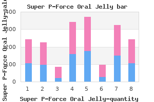
Roberts and colleagues discovered a 24% rate of ureteral stricture following endoscopic therapy of stones fastened in the identical location for greater than 2 months [7] erectile dysfunction treatment with fruits generic 160 mg super p-force oral jelly. Four randomized trials that included proximal ureteral stones had been analyzed by Matlaga et al erectile dysfunction age 22 order discount super p-force oral jelly. Unlike proximal ureteral stones impotence cure 160mg super p-force oral jelly with mastercard, these findings were consistent across stone measurement erectile dysfunction desensitization 160mg super p-force oral jelly with amex. Antegrade ureteroscopy should be thought of in patients with a large stone impacted in the higher ureter erectile dysfunction doctors in louisville ky discount super p-force oral jelly amex. Other high-risk scientific conditions when therapy of renal calculi is inspired are sufferers with a solitary kidney erectile dysfunction young age super p-force oral jelly 160 mg free shipping, reconstructed urinary tract, immunodeficiency, high-risk occupations, poor medical access or compliance, and kids [14]. We also see renal calculi in elderly sufferers and those with significant medical co-morbidities, when the risks of treatment might outweigh any potential profit. All patients with stones bigger than 15 mm demonstrated disease development, 71% with development, 57% ache, and 26% requiring intervention. Some surgeons might perform atrophic nephrolithotomy for these patients with full staghorn calculi who require reconstruction of stenotic infundibula, although these are more and more uncommon [20]. Shock wave lithotripsy is handiest for smaller, non-lower pole, renal calculi. As the stone dimension will increase past 1 cm, ureteroscopic treatment must be favored. However, with higher ureteroscopy experience, more outcomes are being reported of successful ureteroscopic remedy of these bigger stones. Overall, these encouraging leads to chosen sufferers with very massive renal calculi support ureteroscopy as a viable different to the more invasive percutaneous remedy of those patients. Another advantage of ureteroscopic holmium laser lithotripsy is the flexibility to reposition lower pole stones into the higher kidney to enable easier laser lithotripsy and extra profitable residual fragment passage. Stone composition Shock wave lithotripsy shall be less profitable for cystine, calcium oxalate monohydrate, and brushite stones due to their resistance to fragmentation. Relationship of spontaneous passage of ureteral calculi to stone size and placement as revealed by unenhanced helical Ct. Flexible ureteroscopy and laser lithotripsy for single intrarenal stones 2 cm or greater is this the brand new frontier? Comparison of percutaneous nephrolithotomy and retrograde flexible nephrolithotripsy for the management of 24 cm stones: a matched-pair evaluation. Lower pole i: A prospective randomized trial of extracorporeal shock wave lithotripsy and percutaneous nephrostolithotomy for decrease pole nephrolithiasis initial results. Predicting effectiveness of extracorporeal shockwave lithotripsy by stone attenuation value. Renal stone evaluation with dual-energy multidetector Ct and advanced postprocessing strategies: improved characterization of renal stone composition pilot study 1. Prospective long-term followup of sufferers with asymptomatic decrease pole caliceal stones. Additional advances in imaging, together with multidetector Ct scanning, dual-energy Ct scanning, improved sonographic gear and scanning techniques, have additional widened the utilization of imaging in stone illness. Once the analysis of urolithiasis has been made, imaging offers anatomical, functional and physiological information about the stone and the accumulating system, factors that assist in managing therapeutic strategies. Stones within the ureter are normally treated via medical expulsive therapy, hydration, and pain management. Since the majority of urinary tract stones include calcium, most stones that are sufficiently giant (at least 2. Another limitation of plain movie radiography is the dearth of sentimental tissue detail of the viscera, limiting analysis of the kidney, peri- and pararenal fascia, ureter, and so on. As background, in urinary tract obstruction, pathophysiological adjustments affecting the strain in the amassing system and kidney perfusion happen. Ultrasound may be very sensitive for the detection of accumulating system dilation, but the collecting system could also be dilated with out obstruction. A renal or ureteral stone is seen as a filling defect throughout the accumulating system on this modality. Non-contrast Ct was first described as useful within the investigation of stones in 1995, since when it has been repeatedly confirmed to have unparalleled accuracy in the prognosis of urinary tract stones, with a reported sensitivity of 9598% and a specificity of 96100% [3,four,26,27,28,29,30]. However, since 1998, when the primary multidetector (also termed helical or spiral) Ct scanners were introduced, almost all single-slice scanners have been changed by multidetector scanners, starting from two detectors to 128 detectors. Additionally, there have been advances within the postprocessing algorithms and workstations which generate multiplanar datasets. Multidetector attenuation values thus enable for improved differentiation between calcium and uric acid stones utilizing Hounsfield models. Summary imaging of renal calculi is necessary to provide information about the presence of stones, their size and placement, and to depict any related complications. Enteric hyperoxaluria, nephrolithiasis, and oxalate nephropathy: doubtlessly severe and unappreciated problems of Roux-en-y gastric bypass. Prospective evaluation of interobserver variability of the hydronephrosis index and the renal resistive index as sonographic examination methods for the evaluation of acute hydronephrosis. A comparison of noncontrast computerized tomography with excretory urography within the assessment of acute flank pain. Urinary tract stones Part i: position of radiological imaging in analysis and therapy planning. Acute ureterolithiasis: incidence of secondary indicators on unenhanced helical Ct and influence on affected person administration. Ureteral calculi: diagnostic efficacy of helical Ct and implications for remedy of sufferers. Unenhanced helical Ct of ureteral stones: incidence of related urinary tract findings. Value of automated coronal reformations from 64-section multidetector row computerized tomography in the analysis of urinary stone illness. Noninvasive differentiation of uric acid versus non-uric acid kidney stones utilizing dual-energy Ct. Retrograde catheters tend to have smaller lumens than nephrostomy tubes, 67 F being generally employed. However, there seems to be little clear evidence in the literature as to which method of upper tract drainage is to be preferred. One nephrostomy tube was dislodged after the affected person had recovered from sepsis and been discharged from hospital, but before definitive therapy of their stone was undertaken. Ureteral catheterization was not possible in four male sufferers, in two instances because of incapability to tolerate the process and in two because of prostatic enlargement. Of all hospital admissions for therapy of higher urinary tract calculi, 12% required drainage for urinary tract sepsis. Once a affected person has recovered from renal dysfunction or sepsis, they are going to be left to handle with a nephrostomy or ureteral stent whereas awaiting stone passage or definitive management. Studies comparing high quality of life between these with ureteral stents and those with nephrostomies inserted for stone illness have proven no significant difference between the two [21]. Of the 113,459 sufferers who had urgent drainage of the upper urinary tract for infection and urolithiasis, 87. While it could be safe to resuscitate a patient in renal failure overnight and drain their kidney during working hours, particularly if superior to the purpose the place dialysis is required in any case, an infected hydronephrosis requires instant drainage. A small volume of distinction medium could additionally be injected to affirm position on fluoroscopy if required. Sequential dilation of the track is carried out with fascial dilators till a 10 F pigtail locking nephrostomy tube could be placed. Emergency Urinary drainage methods 163 Formal nephrostogram and antegrade placement of a ureteral stent is delayed for 48 h or until the affected person is steady and afebrile. Less generally, a nephrostomy is maintained to enable for percutaneous nephrolithotomy and/or antegrade ureteroscopy. Other complicating factors embody anatomical variants similar to horseshoe or pelvic kidneys. We have a tendency not to perform a retrograde pyelogram within the setting of sepsis provided the guidewire passes simply and into an applicable place on fluoroscopy. Once a guidewire is securely placed, a comparatively large-caliber (8 F) ureteral catheter is superior over the guidewire above the obstruction into the renal pelvis and the guidewire removed to permit for drainage. Shorter stents are inadvisable if the renal pelvis and upper ureter are notably dilated. Placement of a stent may be complicated as a end result of impacted stones or tortuosity of the ureter. An extra-stiff guidewire is sometimes needed to straighten out the ureter and permit for the stent to pass an impacted stone. Single centre evaluation of radiologically-guided percutaneous nephrostomies: a report of 273 procedures. Clinical use of long-term indwelling silicone rubber ureteral splints inserted cystoscopically. Percutaneous nephrostomy and ureteric stent insertion for acute renal deobstruction. Although the therapy of most lower tract stones is easy, there are some challenging clinical situations that can be extra effectually managed with the help of some "ideas and methods. Endoscopic methods and ideas used to handle upper tract calculi may additionally be utilized to the decrease tract in many cases. For instance, entry to the bladder or a continent urinary diversion could also be limited for anatomical causes. At high charges (>15/min), the fiber turns into a "scalpel" and the operator should be succesful of shortly disengage the laser should visualization all of a sudden diminish (irrigant runs out) or the affected person strikes due to gentle anesthesia. While the laser will certainly fragment these "eggshells," a rigid grasper (lithotrite of sorts) can be used to rapidly crush the flakes into small items which are rapidly irrigated. Spinal twine harm For a wide range of reasons, the spinal cord-injured patient is at elevated threat of each higher and decrease urinary tract calculi [12,thirteen,14]. Many of those sufferers are managed by intermittent catheterization or indwelling catheters which are regularly associated with lively urinary an infection or bacterial colonization. Special situations Trauma Occasionally, months following traumatic rupture of the bladder because of exterior violence and pelvic fracture, bone fragments, spicules, and even orthopedic hardware will penetrate the bladder wall and both type extra stones or be taken for a bladder calculus. Unusual displays there are tons of uncommon circumstances which were associated with lower tract calculi, including erosions of an intrauterine system [18], calcified tumors including sarcomas [19,20], encrusting cystitis from Corynebacterium [21], and a lot of self-inflicted overseas physique devices in the psychiatric patient [8]. Prostatic urethral calculi Prostatic calculi true prostatic calculi form throughout the prostate tissue and are usually small and by the way found on prostate imaging or at the time of transurethral prostate surgery. Endoscopic administration is the best method however may require several procedures to eradicate the stones. Post radiation transurethral resection or laser vaporization following radiation therapy for prostate cancer could lead to dystrophic calcification throughout the prostatic urethra. Electroresection, staying inside the lumen of the stent, may be accomplished to clear the tissue ingrowth. Once the scar is cleared, nevertheless, the holmium laser is a wonderful device to fragment any stones in addition to remove the portions of the stent that protrude from the urothelium so as to minimize stone reformation. Flexible scopes can additionally be used, including adult and pediatric sizes if necessary. A grasper and rigid cystoscope (or resectoscope sheath) can be utilized to interact the stent items and remove them. New gadgets With growing innovation, various devices have been used within the urethra. Stone formation has not been reported however the system is still being studied in scientific trials [24,25,26]. Urethral reconstruction Stone formation following urethral reconstruction could occur as a end result of obstruction, stasis, and/or infection. Care have to be taken not to injure the reconstructed urethra resulting within the want for a more sophisticated urethroplasty sooner or later. Summary the overwhelming majority of lower tract calculi may be managed endoscopically due to improved endoscopic access methods and in large part using holmium laser know-how, which can fragment all stones with minimal harm to surrounding tissues. Where obtainable, it has largely replaced the other modalities used in the past to treat lower tract calculi. Ct findings in urinary diversion after radical cystectomy: postsurgical anatomy and problems. Percutaneous nephrolithotomy and cystolithalapaxy for a "forgotten" stent in a transplant kidney: case report and literature evaluation. Erosion of an intrauterine contraceptive system by way of the bladder wall causing calculus: administration and review of the literature. Primary osteosarcoma of bladder diverticulum mimicking intradiverticular calculus: a case report. Prostatic urethral raise: two year results after therapy for lower urinary tract symptoms secondary to benign prostatic hyperplasia. Early retrospective studies have established that ureteroscopy can be safely performed in prepubertal youngsters, reaching related treatment efficacy to that of adults [2,three,4]. Such success has not solely proven ureteroscopy to be feasible but has lead to it being defined because the remedy of selection for youngsters with distal ureteral stones [6,7]. However, the pendulum of stone administration seems to be shifting in the direction of expanding use for ureteroscopy in pediatric stone administration. Singh and colleagues reported their experience with eight kids using ureteral entry sheaths for stone therapy [19]. Furthermore, some of the studies beforehand mentioned embody people older than 18 years of age. When advancing the flexible ureteroscope into the ureter or passing wires and/or lasers via the working port, the scope have to be in a straight and undeflected position to prevent injury to the scope.
Safe 160 mg super p-force oral jelly. Treat for Men's Erection Issues - Coconut Water For Sexual Health.